Creative Biolabs offers a variety of fluorescent dye labeling services for worldwide customers for both research and commercial use. Based on years of conjugation experience, our custom fluorescent antibody and protein labeling service includes experimental design and development of protocols for the preparation of conjugates to be used in various detection systems.
Fluorescent dye labeling, often known as fluorescent tag or fluorescent probe, is the use of chemically bound molecules to detect biomolecules such as proteins, antibodies, amino acids, or nucleic acids. Most importantly, the assays can be performed in real-time, in solution, with high-time resolution and sensitivity down to single molecules. Moreover, the advantages of using a fluorescently labeled antibody include harmlessness to target molecule folding and function, brighter signal, multiplexing capabilities, and ease of use (many are available pre-conjugated to many different colors of dyes).
 Fig.1 Illustration of methods used for fluorescence labeling.1,4
With extensive experience and professional scientists, Creative Biolabs can conjugate your antibody or protein to numerous fluorescent dyes. To verify that the observed fluorescence is a result of staining rather than an unspecific artifact, our skilled staff will work closely with you to help select the most suited dye and labeling site according to your specific requirements. Starting from customer-supplied antibodies or Creative Biolabs generated custom antibodies, we deliver fluorescent dyes labeled antibodies in a relatively short duration. Furthermore, each labeled service is meticulously monitored by our quality assurance center. The fluorescent dyes we can offer are shown below, which include but are not limited to:
Creative Biolabs offers an extensive range of fluorescent dye labeling services for global researchers in the most high-quality and cost-effective way. If you are interested in how we can help you with the development of your programs, please feel free to contact us and get more detailed information.
1. Fluorescent Probe for Optical Fluorescence Imaging of Native Proteins
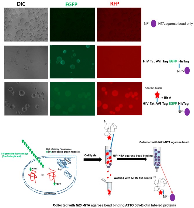 Fig.2 Detection of the Fluorescently Labeled NPI.2,4
This study showcased a highly sensitive fluorescence imaging technique for visualizing a single native protein of interest (NPI), utilizing a eukaryotic translation system that incorporated a cell-permeable fluorescent dye with a free carboxyl group. In living cells, the carboxyl group of the dye interacted with lysine residues on acceptor peptides, and the genetically encoded recognition system achieved nearly 100% fluorescence labeling efficiency. Nickel-nitrilotriacetic acid (Ni-NTA) beads efficiently bound the labeled NPI for detection directly in cells, without the need for purification. The labeling strategy met key criteria for fluorescent NPI measurement using universal carboxyl fluorescent dyes. This approach holds promise for investigating complex biological and ecological processes, enabling single-molecule analysis, and ultra-sensitive NPI detection in nanotechnology applications.
2. Fluorescent Dyes Labeling in Nanoscale Flow Cytometry
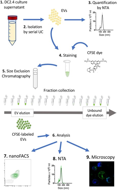 Fig.3 Working strategy of the methods described in this study.3,4
Emerging high-resolution cytometric methods have increased the need for efficient fluorescent labeling techniques to visualize and detect extracellular vesicles (EVs). These labels must be bright enough to detect individual EVs without generating artifacts. This study introduced nanoFACS, a high-resolution flow cytometry method that combined light scattering, fluorescence parameters, and sample enumeration to assess various labels. The approach efficiently stained single EVs, maximizing the fluorescence signal-to-background ratio while preserving functional EV properties. Specifically, researchers compared lipid-, protein-, and RNA-based staining methods and developed this strategy using the amine-reactive fluorescent label 5-(and-6)-Carboxyfluorescein Diacetate Succinimidyl Ester and size exclusion chromatography to remove excess label.
References
For Research Use Only.

