Drawing upon years of experience in merging particles and antibodies, Creative Biolabs extends globally exceptional, efficient, and cost-effective particle coupling services, tailored to specific needs. Our repertoire includes various particle couplings-colloidal gold, colored or uncolored latex, fluorescent latex, nanoparticles, and magnetic particles, among others. These services are available for inclusion in IVD project development or as independent, standalone offerings.
Microspheres stand as fundamental raw materials crucial to the technology employed in protein and nucleic acid detection analysis. Their pivotal role spans a wide array of fundamental fields due to their distinct sizes and specific structures. With varied functions contingent upon particle size and morphology, microspheres play a diverse role. The evolution of detection methods based on microspheres over recent decades has established them as a pivotal tool in IVD and life sciences.
 Fig.1 Magnetic antibody-microparticle conjugation.1,4
In accordance with microsphere structure, categorization reveals three distinct types: porous microspheres, double-layer microspheres, and magnetic microspheres. Presently, diverse microspheres find extensive applications across a spectrum of fields, including biomedicine, cytology, and separation engineering. Noteworthy microsphere properties encompass stability, biocompatibility, functional group attributes, surface characteristics, volume effects, and more.
| Porous Microspheres |
| There are many kinds of porous microspheres, including: polystyrene microspheres, polyurethane microspheres, glass microspheres, ceramic microspheres, etc. Different kinds of porous microspheres have different characteristics. The adsorption of porous microspheres has been widely used in medicine and environmental protection. |
| Double-layer Microspheres |
| Double-layer microspheres with core-shell structure play an important role in the field of medicine and biomedical engineering because of their special structure and variable properties. For example, in pharmaceutical applications, a biodegradable polymer material is mixed with a drug as a microsphere core, and another biodegradable polymer material is a shell. |
| Magnetic Microspheres |
| Magnetic microspheres can not only be endowed with surface functional groups (such as -OH, -COOH, -CHO, -NH2, etc.) by copolymerization and surface modification, but also have guiding functions under the action of external magnetic fields, which are widely used in many fields such as biomedicine, cytology, and separation engineering. |
Creative Biolabs can provide a number of particle conjugation services for in vivo biomedical imaging, targeted drug delivery, and many other applications, which include but are not limited to:
| Colloidal gold | Colored or uncolored latex | Fluorescent latex |
| Nanoparticles | Magnetic particles | Porous carbon microspheres |
| polymeric microsphere | Natural polymer microspheres | SiO2 microspheres |
Our tailored particle conjugation service combines flexibility, reproducibility, and quality assurance, leveraging advanced technology and the expertise of seasoned scientists. For detailed information about how we can fulfill your project requirements, please contact us.
1. Magnetoelectric Nanoparticles for Targeted and Controlled Cancer Drug Delivery and Release
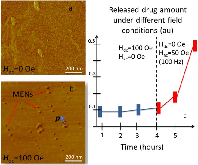 Fig.2 Cancer cell lysate content as a function of ac/dc-field application.2,4
Researchers developed magnetoelectric nanoparticles (MENs) to enhance anticancer drug targeted delivery and release while minimizing damage to normal cells. MENs leverage the electric properties of cancer cell membranes, which require a lower threshold field to induce electroporation compared to normal cells. In vitro and in vivo assays demonstrated that drugs could be attached to MENs via surface functionalization, preventing premature release. When a direct current (d.c.) field was applied, the drug-loaded MENs were directed to cancer cells. Once inside, an alternating current (a.c.) field triggered the controlled release of the drug. By applying a specific sequence of external d.c. and a.c. fields, this method ensured targeted drug delivery to cancer cells, preventing premature loss, enabling selective entry, and on-demand release, while sparing normal tissues.
2. Multifunctional Nanomaterials for Targeted Drug Delivery and Biomedical Imaging
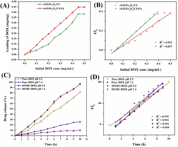 Fig.3 Drug loading and pH-regulated release properties of the rGO/Fe3O4/CS nanocomposite.3,4
This study developed a multifunctional nanosystem for simultaneous cancer imaging and drug delivery. The system was based on a chitosan (CS) polymer functionalized with reduced graphene oxide (rGO) and embedded with Fe3O4 nanoparticles. Its physicochemical properties were characterized using XRD, FT-IR, HR-TEM, FE-SEM, XPS, and VSM analysis. In vivo toxicity studies in zebrafish indicated that the nanocomposite was non-toxic. The system demonstrated a drug loading capacity of 0.448 mg/mL for a chemotherapeutic agent, a common anticancer drug. pH-triggered release was facilitated using folic acid as a targeting ligand. Cellular uptake and multimodal imaging showed that the folic acid-conjugated nanocomposites significantly increased drug delivery to folate receptor-positive cancer cells. Additionally, the nanocomposite exhibited improved antibiofilm and antioxidant properties in comparison to other materials. These findings highlight the nanocomposite's potential in targeted chemotherapy and applications in polymer, biomedical, cosmetic, etc.
References
For Research Use Only.

