B Cell-based Phage Display Assay
Creative Biolabs has developed a robust phage display platform for B-cell epitope mapping and provides economic and rapid approaches to cancer epitope analysis.
B Cell-based Phage Display Assay
Phage display is a high-throughput technique that can display billions of foreign peptides on bacteriophage through the fusing of target peptide genes into coat genes. This approach allows the expression of proteins on the surface of phage in a multivalent type of short peptides or limited copies of long peptides.
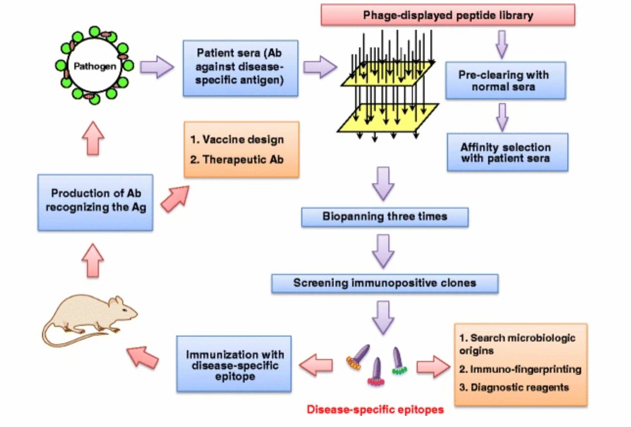
Epitope is the immunogenic determinant of an antigen that is recognized by T cells, B cells, and antibodies. In previous studies, a random peptide library displayed on phage was adopted to screen B cell epitopes using monoclonal antibodies and polyclonal antibodies immobilized on a support plate or bound on magnetic beads.
 Fig.1 Using phage-displayed peptide libraries and biopanning selection to identify antigen-specific epitopes. 1
Phage Display-based Cancer Epitope Analysis at Creative Biolabs
Creative Biolabs applies phage display technology to select high-affinity antigenic peptides of an antigen protein from billions of candidates displayed on phage in a recombinant expression library. Subsequently, the candidate epitopes can be further screened and enriched with a highly selective biopanning approach.
Workflow

Phage Displayed Peptide Library Construction
-
Full-Length Antigen Proteins Acquisition. We use the DNA sequences of the full-length antigen proteins to identify specific immunogenic epitopes. The DNA sequence information can be derived from whole-genome sequencing and RNA-seq of patient biopsies tumor tissue.
-
Peptides Library Synthesis. A large-scale, random, 12-mer peptide library is generated using trinucleotide oligos to avoid stop codons and use amino acid native-like.
-
Phage Display. The peptide library is inserted into E. coli via the engineered protein scaffold.
Potential Antigen Epitope Biopanning
-
Antibody Preparation. Dilute serum samples obtained from patients or immunized mice.
-
Antibody Incubation. Antibodies are added to plates containing combinational library cells and incubated for hours.
-
Reactive Epitope Separation. Using magnetic beads bound with protein A/G to capture human/mice serum antibodies interacted with phage-displayed epitopes.
-
Cancer-Specific Epitope Identification.
-
Magnetic separation system and FACs analysis
-
Next-generation sequencing
-
Several rounds of magnetic affinity separation
Highlights
-
High-throughput Assay
-
Genome-scale Screening
-
High-efficient and Reliable Approach
Get Access to Our Services
Creative Biolabs is devoted to developing comprehensive, integrated services covering the broad application of phage display technology. We are devoted to providing high-quality antibody and antigen phage display and customized screening services to meet our partner's particular needs and objectives. Please contact us for further information.
Reference
-
Wu, C. H.; et al. Advancement and applications of peptide phage display technology in biomedical science. Journal of biomedical science. 2016, 23, 8.
For Research Use Only | Not For Clinical Use


 Fig.1 Using phage-displayed peptide libraries and biopanning selection to identify antigen-specific epitopes. 1


 Download our brochure