ELISpot Assay Service
Let Our Experienced Team Help You With the ELISpot Assay
Equipped with a professional team and a variety of cutting-edge technologies in ELSIPOT, Creative Biolabs seeks to facilitate the advancement of research in acquired immunodeficiency syndrome (AIDS), cancer, infectious diseases, and autoimmune diseases.
ELISPOT is a good choice to detect cytokines spontaneously, such as IFN-gamma, TNF-alpha, IL-2, and IL-4, which are produced by peripheral blood lymphocytes. To measure antigen-specific T cells, which typically occur at low frequency in vivo, ELISpot techniques also are highly sensitive functional assays. Creative Biolabs offers ELISpot assays using commercially available or custom assays to support our clients' R&D projects, and we provide validation services. Having performed ELISpot for over 10 years, we offer a series of tips to maximize the success of your test. There are 6 general steps of this assay, as shown below.
 Fig.1 6 General steps of ELISPOT.
B Cell ELISPOT Assays and T Cell ELISPOT Assays
We are proficient at performing B-cell ELISPOT and T-cell ELISPOT assays. The ELIspot can provide valuable information about the amount of cytokine-producing cells for CD4+ T cells and CD8+ T cells, while for B cells, we also determine the specific isotype of produced antibodies. Our scientists are experienced in performing ELISpot analysis for a single cytokine or a combination of two or more cytokines. We have developed various customized B-cell ELISPOT and T-cell ELISPOT assays for customers in the field of pharmaceutical and biotechnology.
Below is the Workflow of the Antigen-Specific T Cell ELISpot Assay:
-
Peripheral blood mononuclear cells (PBMCs) or other tissue cells are cultured on a membrane surface with or without antigenic stimuli. The membrane surface is precoated with a specific capture antibody with or without antigenic stimuli.
-
The cytokines secreted by cells are absorbed on the membrane surface.
-
After incubation, cells are removed. Then, the secreted cytokines are measured using a second detection antibody.
The spots are visible because of the detection antibody conjugated with alkaline phosphatase or horseradish peroxidase (HRP).
-
Then, wash the plate and stop the reaction.
-
Analyze the results before the membrane is dry.
Advantages of ELISpot Assays
-
Optimized, high-throughput assays
-
Highly flexible: ELISpot can be applied to multiple readout formats.
-
Quantitative: The key cellular functions of immune cells can be measured by ELISPOT.
-
Both adaptive and innate immune responses can be assessed by ELISpot.
-
Using common standard operating procedures, the ELISpot assay can be standardized, resulting in the translation of the assay from the laboratory to a preclinical or clinical tool.
Applications of ELISpot Assays
-
Measurement of the immune response to infection by B cell ELISpot assays
-
Qualification of antigen-specific antibody-secreting cells in human PBMCs
-
Assessment of next-generation therapeutics
-
ELISpot analysis can be used in multiple research areas including allergy, infectious diseases, cancer, vaccination studies, and autoimmunity.
Publications Sharing Based on ELISpot Assays
Publication 1
Cells Used: Tumor-infiltrating lymphocytes (TILs)
Journal: Journal of Investigative Dermatology
IF: 6.5
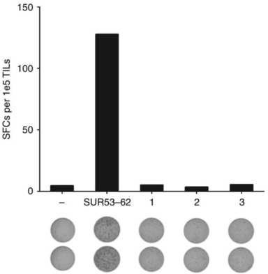
Research Findings: ELISPOT assay represents one of the central methods in clinical diagnostic settings for the investigation and characterization of antigen-specific T cells, for example, in the context of tracking immune responses to vaccines.
 Fig.2 IFN-γ ELISPOT assay.1
For more information and a detailed quote, please contact us.
Reference
-
Möbs.; et al. "Research techniques made simple: monitoring of T-cell subsets using the ELISPOT assay." Journal of Investigative Dermatology 136.6 (2016): e55-e59.
For Research Use Only | Not For Clinical Use


 Fig.1 6 General steps of ELISPOT.
 Fig.2 IFN-γ ELISPOT assay.1
 Download our brochure