Raman Imaging Service for In Situ Organelle Metabolic Research
Visualize the Responses of Metabolites Precisely
Raman microspectroscopy, a spectroscopic technique that uses the inelastic scattering of monochromatic light to allow direct chemical observation of biomolecules like DNA and RNA, proteins and lipids in uncloaked cells and tissues, does not require labeling the molecules concerned. For precise analysis of signals from molecules present in specific organelles of cells, such as proteins, fatty acids and lipids, DNA, RNA, carbohydrates, carotenoids, retinoids, and primary metabolites, Raman microscopy is an efficient tool. Raman imaging is not only a mere microscopy, it offers information on the chemical composition of brain tissue with lipid-rich, protein-rich, and mixed profiles due to an integrated Raman spectra that are embedded into tissue images.
Applying Raman imaging for tumor subcellular metabolic analysis, Creative Biolabs enables the precise metabolic analysis of cancer cells.
Advantages of Raman Imaging
-
The detection of the molecular vibrations.
-
Reflection of the structures and chemical conditions of molecules.
-
Direct visualization of the chemical responses of endogenous molecules.
Applications of Raman Imaging
-
Analysis of metabolic precursors of biomolecules
-
Provide valuable information on small bioactive molecules, such as natural products and drug candidates
-
Help understand the intracellular localization of molecules
-
Help understand the molecules and their interactions with biomolecules
-
Monitoring the uptake and distribution of drug candidates
Publications Sharing
Publication 1
Method Used: Label-free Raman imaging in cell membranes
Journal: Annual Review of Analytical Chemistry
IF: 8
Research Findings: The cell membrane, which consists of various proteins, lipids, and small molecules with distinctive chemical properties, is an elaborate organelle. This paper described the use of these methods to image membrane proteins and lipids, concluding that a labelless Raman imaging technique is an effective method for cell analysis.
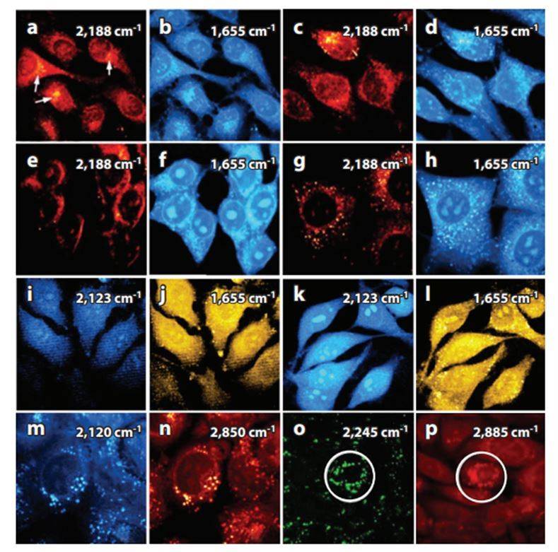 Fig.1 Stimulated Raman scattering images.1
Publication 2
Method Used: Raman imaging for monitoring intracellular retinoid metabolism in cancer cells
Journal: Journal of Molecular Liquids
IF: 6
Research Findings: The authors have developed a method to detect molecular processes taking place in normal human brain cancer cells due to the transport of retinol at isolated organelles levels, by using label-free Raman techniques for whole-cell biochemical imaging. The results have shown that Raman imaging, which can detect a separate spectrum signature of vibrations, has been an efficient tool for the assessment of retinoids and Retinol-bound proteins associated with carcinogenesis.
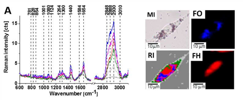 Fig.2 Confocal Raman spectroscopy analysis of the glioblastoma (U-87 MG) cells.2
At Creative Biolabs, we provide comprehensive Tumor Metabolism Characterization Services including but not limited to:
Please contact us if you would like to discuss any of these services.
References
-
Syed, Aleem, and Emily A. Smith. "Raman imaging in cell membranes, lipid-rich organelles, and lipid bilayers." Annual Review of Analytical Chemistry 10 (2017): 271-291.
-
Abramczyk, Halina, Anna Imiela, and Jakub Surmacki. "Novel strategies of Raman imaging for monitoring intracellular retinoid metabolism in cancer cells." Journal of Molecular Liquids 334 (2021): 116033.
For Research Use Only | Not For Clinical Use
 Download our brochure