B Cell-based Quenching Assay
Summary of Quenching Assay
An appropriate method that can be used to monitor the immune responses between the antibody and antigen peptides is valuable for immunogenic epitope discovery. Creative Biolabs offers a beads-based immunoassay to identify epitopes that can bind to specific antibodies. The candidate peptides are conjugated on beads and incubated with positive antibodies. Fluorescent-labeled anti-antibody and different lasers combined to detect the Ab: Ag complex formed on the beads. Creative Biolabs provides a set of beads that is coded with unique labels and has a discriminate spectrum to each other and tailored cancer epitope analysis services using this convenient and moderate throughput beads-based assay.
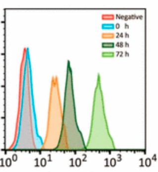
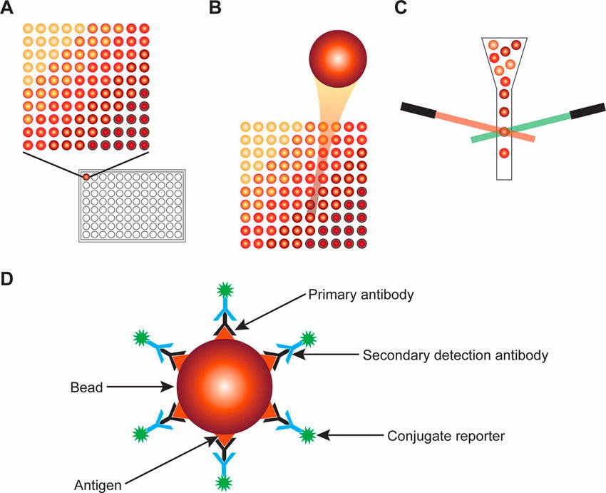 Fig.1 Using the laser quenching method detects specific fluorescent-antibodies bound antigen epitope on the surface of the beads.2
Workflow
 Fig.2 Workflow (Creative Biolabs)
Key Features of the Service
 Fig.3 Key Features (Creative Biolabs)
Our Capabilities

Complementary Validation Assays
|
Complementary Assays
|
Detailed Description
|
|
Dose-dependent Ab:Ag Reactive
|
Immunized plasma samples are diluted to multiple doses to monitor dose-related reactions to epitope peptides.
|
|
Peptide-specific ELISA
|
The specific antibody absorption response to each epitope is confirmed using ELISA.
|
|
Peptide-induced T-cell Responses
|
Peptides are expressed on cell lines and inoculated with immunized PBMC to test
IFN-r production, cytotoxicity, and competitive antibody-based inhibition test.
|
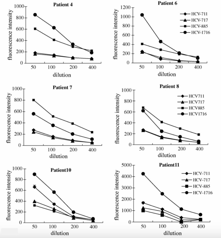 Fig.4 IgG levels of patient-diluted plasma to four identified HCV epitopes.1
Contact Us
At Creative Biolabs, you just need to tell us what target antigen you are interested in. Our experienced in-house experts will provide comprehensive solutions to investigate immunogenic epitopes in the antigen and discover novel candidate epitopes. Please contact us and get the protocol for quenching assay.
Related Services
References
-
Tamura, M.; et al. Identification of hepatitis C virus 1b-derived peptides recognized by both cellular and humoral immune systems in HCV1b(+) HLA-A24(+) patients. Hepatology research : the official journal of the Japan Society of Hepatology. 2005, 32(4): 227–234.
-
Ragan, I. K.; et al. Evaluation of fluorescence microsphere immunoassay for detection of antibodies to rift valley fever virus nucleocapsid protein and glycoproteins. Journal of Clinical Microbiology. 2018, 56(6): 10-1128.
-
Cartechini, L.; et al. Immunochemical Methods Applied to Art-Historical Materials: Identification and Localization of Proteins by ELISA and IFM. Topics in Current Chemistry. 2016, 374.
-
Musicò, A.; et al. SARS-CoV-2 Epitope Mapping on Microarrays Highlights Strong Immune-Response to N Protein Region. Vaccines. 2021, 9(1): 35.
-
Zhao, Y.; et al. Single cell RNA expression analysis using flow cytometry based on specific probe ligation and rolling circle amplification. ACS sensors. 2020, 5(10): 3031-3036.
For Research Use Only | Not For Clinical Use


 Fig.1 Using the laser quenching method detects specific fluorescent-antibodies bound antigen epitope on the surface of the beads.2
 Fig.2 Workflow (Creative Biolabs)
 Fig.3 Key Features (Creative Biolabs)

 Fig.4 IgG levels of patient-diluted plasma to four identified HCV epitopes.1
 Download our brochure