Glioblastoma (GBM) is one of the most aggressive brain tumors, characterized by rapid proliferation, invasive behavior, and poor prognosis, with average survival limited to 12-15 months after diagnosis. One of the greatest challenges in studying GBM is the tumor's ability to infiltrate healthy brain regions, making it difficult to assess through conventional in vitro models, such as two-dimensional (2D) cell culture assays. While 2D assays are widely used for tumor studies, they fail to capture the complex interplay between tumor cells and the central nervous system (CNS) microenvironment.
Creative Biolabs has addressed these limitations through our ex vivo human brain slice assay, with a cutting-edge platform that utilizes living neuronal tissue to closely mimic the natural brain microenvironment. This method enables precise evaluation of GBM progression, offering invaluable insights into the interactions between tumors and their host tissues. The ex vivo human brain slice model provides a more realistic alternative to conventional models, accurately replicating the cytoarchitecture and physiological conditions required to advance oncology and neuroscience research.
Get Customized Services
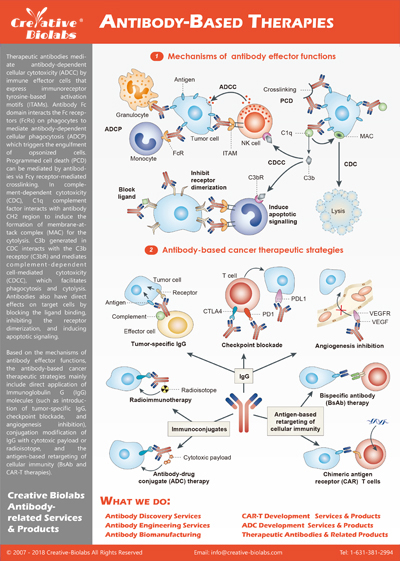
Workflow for Ex Vivo Brain Slice Assay
The ex vivo human brain slice assay requires rigorous preparation and maintenance to ensure the neuronal tissue closely resembles in vivo conditions. Below is a detailed breakdown of the key steps involved in generating and maintaining brain slices for research purposes at Creative Biolabs. More assays (e.g. Rodent Brain Slice Assay) about brain slices can be found in the unit of Organotypic Brain Slice Assays.
1. Brain Tissue Collection and Source
The clients' provided tissue used in this assay should be derived from therapeutically resected human brain samples, which can ensure the highest clinical relevance. Regions such as the neocortex and hippocampus are prioritized due to their significance in neurological and oncological research. These areas provide access to critical neuronal populations, including pyramidal neurons, interneurons, and dentate gyrus granule neurons.
2. Sectioning with Vibratome
Using a vibratome, coronal brain sections are prepared with precise thickness, typically between 350-400 μm. The slicing process is meticulously calibrated to preserve the tissue's cytoarchitecture and maintain essential features such as neuronal connectivity and glial-neuronal interactions. The accuracy of this step ensures consistency across samples, facilitating reproducible and reliable data collection.
3. Transfer and Culturing of Brain Slices
After sectioning, the brain slices are carefully transferred to a nylon membrane insert within a six-well plate using a blunt, fire-polished glass Pasteur pipette to prevent damage.
Media Composition: The slices are cultured in minimal media without excessive growth factors to maintain the endogenous characteristics of the tissue.
Air-Liquid Interface: To ensure optimal viability, the brain slices are maintained at an air-liquid interface, preventing full immersion in the medium while allowing nutrient absorption and gas exchange.
4. Brain Slice Characterization and Validation
The functional integrity and viability of the brain slices are confirmed using several advanced characterization methods:
5. Spheroid Implantation (Optional for Tumor Studies)
In neuro-oncology research, tumor spheroids can be implanted into the prepared brain slices to replicate the invasion patterns of GBM. The spheroids are carefully positioned within the tissue to ensure the natural progression of tumor infiltration. Imaging methods such as confocal microscopy and epi-fluorescence imaging are employed to monitor tumor invasion in real time.
6. Experimental Customization and Treatment Protocols
This platform offers flexibility for a variety of experimental designs, including:
Drug Treatment: Brain slices and implanted tumor spheroids can be treated with therapeutic compounds to assess their effects on tumor behavior and neuronal health.
Gene Knockout Models: Brain slices from genetically modified donors are used to investigate the role of specific genes in tumor progression and CNS function.
Why Our Ex Vivo Human Brain Slice Assay Stands Out
At Creative Biolabs, we offer cutting-edge ex vivo human brain slice assays designed to overcome the limitations of conventional models. Our platform provides superior biological relevance, flexibility, and precision, giving researchers the most reliable tools for breakthrough discoveries.
Good Clinical Relevance
Unlike traditional murine models that lack mature brain structures, our assays utilize adult human brain tissue to replicate the exact microenvironment encountered in the central nervous system (CNS). This ensures your research closely mirrors in vivo conditions.
Enhanced Imaging Quality
We employ lipophilic dyes and tissue-clearing techniques, significantly improving imaging clarity and eliminating autofluorescence. This allows for precise observation of tumor invasion and neuron-tumor interactions in real time.
Customizable Experimental Design
Whether you need drug testing, gene knockout models, or co-culture studies, our platform offers unmatched flexibility. We tailor every experiment to your specific research objectives, providing actionable data for oncology, neuroscience, and pharmacotherapy research.
Creative Biolabs offers expertise, in gathering robust data to drive confident decision making throughout your Brain-Immune-Gut Based Integrative Study Research. If you have any questions about your microbe project, please feel free to contact us.


 Brain-Immune-Gut interaction Study
 Download our brochure