T Cell-based CFSE Assay
The findings that each cancer contains many mutant genes do not present in normal tissues have prompted considerable interest in the cancer epitope landscape. Creative Biolabs is dedicated to offering cancer epitope identification, assay development, and validation services to our clients with the most potential candidates to accelerate their drug development.
CFSE-based Proliferation Assay
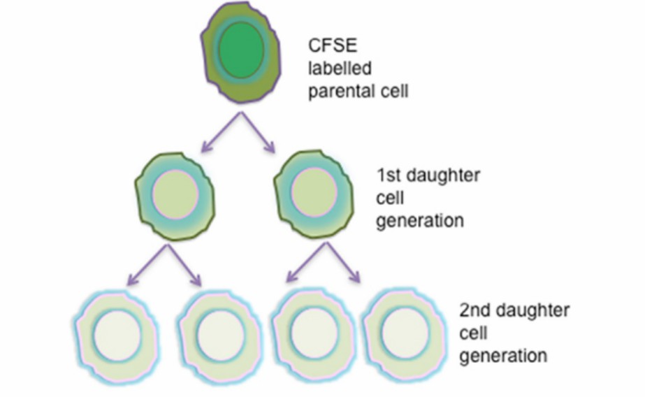
 Fig.1 CFSE proliferation assay.1
Cancer antigens are the consequence of somatic mutations in individual tumors and are not present in normal tissues. The lack of in-depth study of the peptide epitope information of the individual tumors impairs the advance in tumor immunity study.
The specific response of T cells to target tumor cells can be applied to analyzing cancer epitopes. The carboxyfluorescein succinimidyl ester (CFSE) based method is a non-radioactive way to measure cell expansion. When penetrating living cells, it undergoes esterase cleavage and conjugates with intracellular protein covalently, generating fluorescently labeled cells. As cell proliferation, the CFSE is equally divided into progeny cells.
T Cell-based CSFE Assay Services at Creative Biolabs
The CFSE-based fluorescent dye assay has widely been used to study the cytotoxicity of cytotoxic T lymphocytes in vivo. Based on this experiment, Creative Biolabs applies it to T cell-relevant cancer epitope analysis and develops an easy and quick assay. The assay is carried out based on the cytotoxicity of specific T cells on target cells and following the reduction of target cell signals.
 Fig.2 Workflow of T Cell-based CSFE Assay for cancer epitope analysis.3
Description of the Main Steps
 Fig.3 The CSFE-based assay service at Creative Biolabs.
Advantages and Features
-
Compatible Multi-staining
CFSE bleeds over into multiple channels on the flow cytometer and supports co-stain with PeCy7 and other fluorophore-labeled antibodies.
The substance has less or no impact on cell viability and proliferation.
The fluorescent dye is stable and efficiently stains live cells, retaining them for several days.
-
Excellent in Cell Labeling
The CSFE enters cells indiscriminately and diffuses throughout the cytoplasm. The stain results are consistent and uniform between cells.
The results obtained from our assay are consistent with those traditional methods.
Applications of the CSFE Assay
✔ Measure cell division in vitro.
✔ Assess cell kinetics in vivo.
✔ In vivo tracking of stained cells by fluorescent microscopy.
The bioassay platform established by Creative Biolabs is a valuable and validated tool that can be used for in vitro and in vivo analysis to study the T and B cell epitopes of cancer. We offer a wide range of assays for epitope identification, including cell proliferation, cytotoxicity, cytokine release, and T and B Cell-based Binding Assay. Please feel free to contact us or send us a query directly.
References
-
Lokhande, M. U.; et al. Methodologies for the Analysis of HCV-Specific CD4(+) T Cells. Frontiers in immunology. 2015, 6: 57.
-
Durward, Marina.; et al. Antigen specific killing assay using CFSE labeled target cells. J Vis Exp. 2010, (45): 2250.
-
Ganesan, N.; et al. Methods to Assess Proliferation of Stimulated Human Lymphocytes In Vitro: A Narrative Review. Cells. 2023, 12: 386.
For Research Use Only | Not For Clinical Use


 Fig.1 CFSE proliferation assay.1
 Fig.2 Workflow of T Cell-based CSFE Assay for cancer epitope analysis.3
 Fig.3 The CSFE-based assay service at Creative Biolabs.
 Download our brochure