T Cell-based Cytometric Bead Array Assay
The success of the immunotherapy is associated with the epitope-specific T-cell immune responses that are strong and broad. With decades of bioassay developing experience, Creative Biolabs offers the flow cytometric beads-based assay for discovering T cell-associated cancer epitopes.
T Cell-based Cytometric Bead Array Assay
Based on the epitope-specific cytokines release response from T cells, it is possible to pick the epitope candidates that could stimulate robust T-cell responses. Traditionally, enzyme-linked immunosorbent assay (ELISA) is used to quantitatively measure the presence of cytokines in a sample. Cytometric bead assay is a technique that melds ELISA-based technology with flow cytometry to simultaneously test multiple analytes in a small volume of sample in a single reaction.
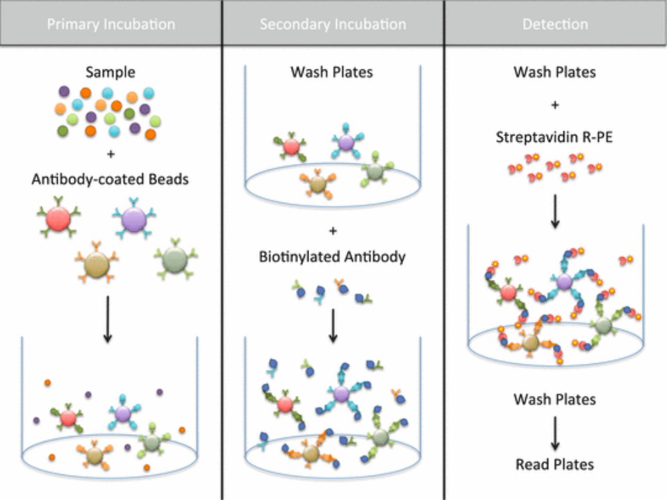
 Fig.1 Quantification of multiple cytokines and chemokines using cytometric bead arrays.1
Cytometric Bead Array (CBA) Service at Creative Biolabs
Creative Biolabs has developed flow cytometric bead-based immunoassays to evaluate epitope-specific cytokines secretion from T cells. In the assay, a mixture of fluorescent-labeled microbeads and highly specific antibodies are designed to capture cytokines, and flow cytometry is utilized to read out the results of the immunoassays.
Workflow

Overview from Sample to Analytics
Sample Types
Plasma
Cell culture supernatant
Sample Volume
25 to 50ul per test
Analytical Space
10 different analytes simultaneously
30 proteins at maximum
Characteristics of the CBA Assay Service
The quantitative limitation of the assay is lower than 1pg/ml of cytokines in samples.
Include assays for ten different cytokines from various Th1/Th2 or inflammatory cytokines that can be combined to develop a multiplexed panel.
-
Save Much Time and Resources
Simultaneously detect multiple analytes from a relatively small volume sample in a rapid approach.
Our assay can analyze up to 10 proteins with just 25 to 50ul of a sample.
Validated High Performance of the Assay
Comprehensive Evaluation
✔ Combined single-plex and multiplex situations to ensure consistency across assay types.
✔ The capture antibody and detection antibody have been evaluated for dynamic range, sensitivity, and dilutions.
Optimized Assay Protocol
✔ A two-step detection protocol to maximumly reduce the background fluorescence and maximize analyte-related signal.
Accurate Data Analysis
✔ The average fluorescence intensity (MFI) is calculated based on the analysis of the fluorescence intensity of each analyte on each bead in the mixed microspheres.
The List of Analytes in the Assay
|
IL-1b
|
IL-6
|
IL-10
|
IL-17A
|
IL-23
|
Granzyme A
|
CCL2/MCP-1
|
CXCL10/IP-10
|
|
IL-2
|
IL-7
|
IL-12
|
IL-17F
|
IFNg
|
Granzyme B
|
CCL4/MIP-1b
|
|
|
IL-4
|
IL-8
|
IL-13
|
IL-21
|
TNF
|
GM-CSF
|
CCL5/RANTES
|
|
|
IL-5
|
IL-9
|
IL-17
|
IL-22
|
TNFa
|
TGFb
|
CXCL9/MIG
|
|
In addition to the analytes above, Creative Biolabs also provides functional beads conjugated with your antibody for the antigen of your interest. We are dedicated to helping our clients meet the various demands of cancer epitope identification. If you are interested in the service, please contact us.
Reference
-
Moncunill, G.; et al. Quantification of multiple cytokines and chemokines using cytometric bead arrays. Methods in molecular biology (Clifton, N.J.). 2014, 1172: 65-86.
For Research Use Only | Not For Clinical Use


 Fig.1 Quantification of multiple cytokines and chemokines using cytometric bead arrays.1

 Download our brochure