B Cell-based Immuno Staining Assay
Principle of the Technique
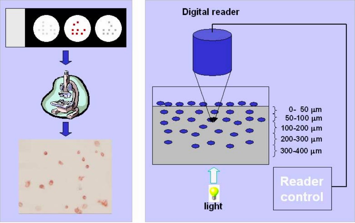
Immunostaining assay is an indispensable immune cytologic technique used to detect various molecules in cells and tissues. This valuable technique is widely used in disease diagnosis, prognosis assessment, and biomarker identification. In an immunostaining assay, ex vivo cell cultures or cells separated from tissues and blood, despite adherent cells or suspension cells, are employed for measurement.
The experiment is done using various chemical reagents for fixation and permeabilization to preserve the protein location and structure state and facilitate antibodies' transportation and binding. Reliable and specific antibodies are the key point of the staining and can be visualized by microscopy.
 Fig.1 Illustration of immuno staining assay.1
Immuno Staining Assay Service at Creative Biolabs
Creative Biolabs offers a validated protocol for immunocytochemistry, including protein synthesis, reactive antibody production, and immuno staining assay. The assay is commonly used for infectious virus binding epitope analysis. The impact of neutralizing antibody addition on virus infectious is detected by immuno staining assay.
Workflow

Protein Synthesis
-
Proteins of antigen are synthesized or overexpressed from recombinant plasmids.
-
The recombinant proteins are purified and validated using affinity chromatography and SDS-PAGE.
Neutralizing Antibody Production
-
Mice are immunized with purified recombinant proteins and Freund's adjuvant in two-week intervals. Neutralizing antibodies are collected from mice sera and analyzed using ELISA.
-
Other animal models, including rabbits, pigs, and non-human primates, are also available.
-
Isolate antigen-specific antibodies from the serum of patients.
 Fig.2 Immuno staining results of a neutralizing monoclonal antibody on HCV infection.2
Protocol of Immuno Staining Assay
-
Host cells are inoculated with the virus with or without antibody pre-incubation.
-
After washing cells with PBS, monolayer cells are fixed with cold methanol or paraformaldehyde.
-
Then, cells are blocked with serum from the same species with the secondary antibody to eliminate non-specific binding.
-
The fixed cells are incubated with the specific diluted antibodies for several hours after a washing step. Cell nuclei are stained with dyes at the same time.
-
Finally, a fluorescent-labeled secondary antibody is inoculated with cells to detect bound antibodies.
-
Monolayer cells are examined by microscope following washing off the unbound antibodies.
Features of the Service
|
Reporter
|
Examples
|
Color
|
Visualization
|
Specificity
|
|
Enzyme
|
Peroxidase-DAB
|
Brown
|
Light-microscopy
|
Antigen
|
|
Fluorophore
|
FITC, Cy3
|
Green, yellow
|
Fluorescence microscope
|
Antigen
|
|
Dyes
|
DAPI
|
Blue
|
UV light
|
Cell nucleus
|
-
Simultaneously detect several antigens in the cell.
-
Subcellular localization of target antigens.
-
Both direct and indirect staining are available with high sensitivity and fast turnaround.
-
High-resolution staining images from confocal laser scanning microscope, digital imaging, and electron microscopy.
Related Service for Epitope Analysis
Immuno staining assays are used to validate the neutralization function of antibodies to viruses, and advanced methods are necessary to analyze crucial epitope peptides in protein. The following assays are recommended to you to find a proper strategy.
Cancer Epitope Analysis Assays at Creative Biolabs
With cutting-edge instruments and expertise, Creative Biolabs is devoted to offering sensitive, reliable, reproducible immuno staining assays for the identification of antigenic epitopes in cancer antigens. If you are interested in our immuno staining assay, please contact us and get a personalized scheme.
References
-
Li, S. S. Thrombospondin 1, an Autocrine Regulator in T Cell Adhesion and Migration. 2023.
-
Yi, C.; et al. Junctional and somatic hypermutation-induced CX4C motif is critical for the recognition of a highly conserved epitope on HCV E2 by a human broadly neutralizing antibody. Cellular & molecular immunology. 2021, 18(3): 675–685.
For Research Use Only | Not For Clinical Use


 Fig.1 Illustration of immuno staining assay.1

 Fig.2 Immuno staining results of a neutralizing monoclonal antibody on HCV infection.2
 Download our brochure