The glucocorticoid receptor (GR) belongs to the nuclear receptor superfamily of intracellular proteins that function as ligand-dependent transcription factors. Upon binding hormone, the receptor enhances or represses expression of target genes leading to specific changes in cellular function. Post-traumatic stress disorder (PTSD) is reported in some studies to be associated with increased GR sensitivity. Creative Biolabs is dedicated to establishing the most exquisite service platform for our clients and the one-stop nuclear receptor modeling services can provide comprehensive technical support to advance our clients’ projects.
The glucocorticoid receptor functions as a transcription factor that binds to glucocorticoid response elements in the promoters of glucocorticoid-responsive genes and activates their transcription. Reduced glucocorticoid signaling was observed before, and shortly after, trauma exposure in those who developed PTSD, which confirms the hypothesis that insufficient glucocorticoid signaling at the time of trauma results in unopposed sympathetic nervous system activation that enhances the consolidation of the traumatic memory. The formation of strong emotional memories is adaptive because remembering danger when appropriately signaled might enhance survival. However, in the absence of sufficient glucocorticoid signaling, the distress that occurs once reminded, and the generalization of triggers, could result in a cascade of maladaptive symptoms.
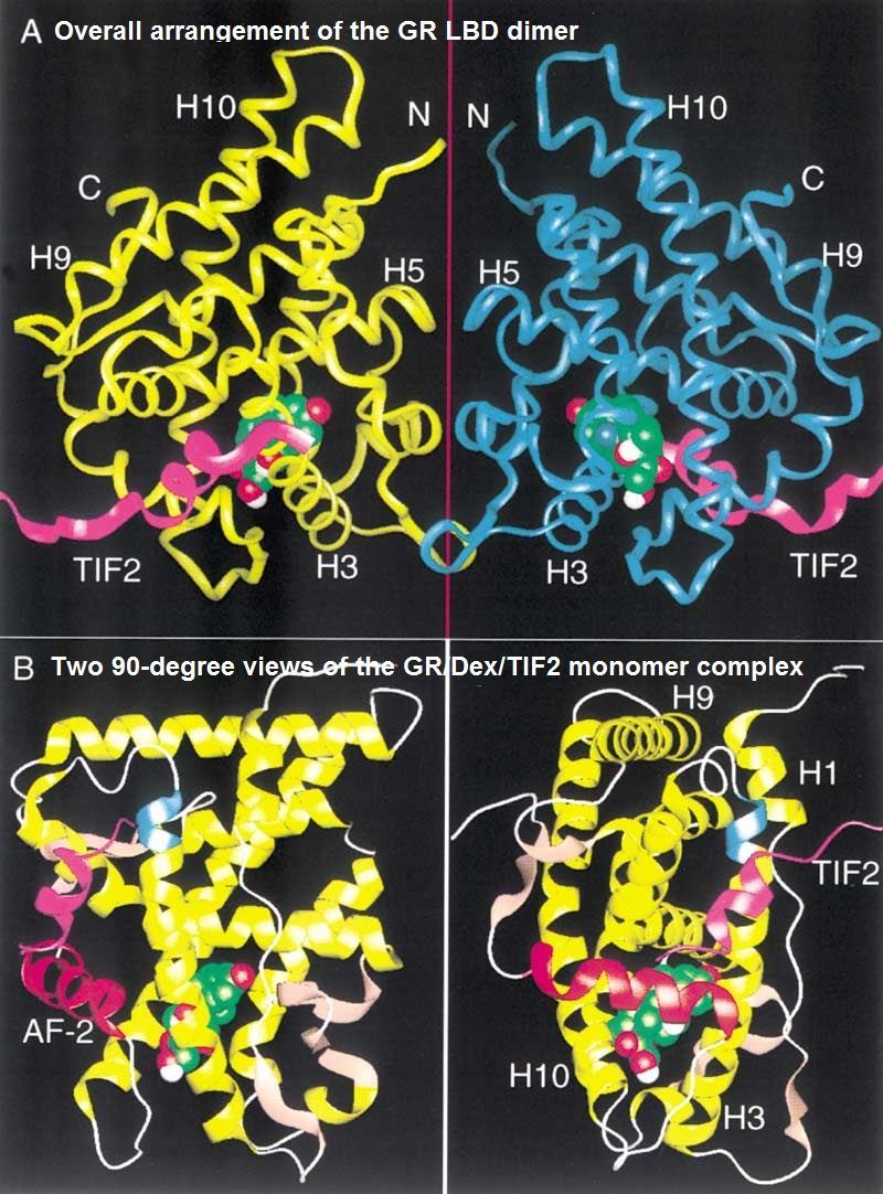
The GR consists of a C-terminal ligand-binding domain (LBD), a DNA-binding domain (DBD) with a dimerization interface in the center of the molecule and two transcription activation (AF) domains. One transcription activation domain (AF1) is located at the N-terminal end of the protein, while the other (AF2) lies within the LBD and is therefore directly controlled by the ligand. The DBD is the most conserved domain across the nuclear receptor family and contains two zinc finger motifs that recognize and bind target DNA sequences. Three amino acids comprising the ‘P-box’ are found in the first zinc finger and are important for target site discrimination, and five amino acids comprising the ‘D-box’ are found in the second zinc finger and are important for receptor dimerization.
 Fig.1 Structure of the GR/Dex/TIF2 complex.
Crystallography of the GR assists the understanding of the structural principles of ligand interactions with their target receptors, but the ultimate goal of computational NR biology is the prediction of affinity, activity and selectivity profiles for novel compounds. For that, the crystal structures need to be converted into predictive models and carefully benchmarked in retrospective applications. The two complementary approaches to the problem address it from the side of the binding pocket and from the side of the co-crystallized ligand. In both cases, the prediction is based on structural modeling and the docking of flexible compounds. As an expert in the structure-based nuclear receptors modeling filed, we can offer a variety of nuclear receptors modeling services to meet specific customers’ requirements.
Creative Biolabs can help accelerate your drug discovery journey towards the clinic and we whole-heartedly cooperate with you to accomplish our shared goals. Our team provides you with outstanding support and meets your specific needs with a professional technology platform. If you are interested in our services, please contact us for more details.
All listed services and products are For Research Use Only. Do Not use in any diagnostic or therapeutic applications.