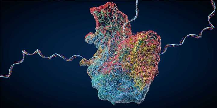
Ribosome display technology was reported more than 20 years ago. Its essence is to utilize the formed mRNA-ribosome-protein trimer complexes to link the nascent proteins to their mRNAs through ribosomes. However, the current ribosome display technology has some drawbacks, such as some proteins that are harmful to the cell. Proteins that play important regulatory functions in the cell cannot be selected, and the diversity of libraries is low. In order to solve the above problems, a new ribosome display technology has been developed, which is the ribosome inactivation display system. The ribosome inactivation display system is considered to be a powerful technology for selecting and evolving peptides and proteins. Based on our rich field experience and ribosome display platform, Creative Biolabs provides comprehensive services to support the development and application of ribosome inactivation display systems.

Display systems can screen DNA or RNA associated with their genotypes from libraries where proteins and peptides are phenotyped, as well as evolve through multiple rounds of mutation, screening, and replication, ultimately altering phenotypic characteristics. Ribosome display, bacterial display, phage display, yeast display, and other surface display technologies have been widely popularized in the past 30 years or so. They play an important role in the field of antibody screening.
Ribosome display technology has some limitations and drawbacks. Because cell-dependent display systems include required in vivo steps, transformation efficiency and protein properties limit the size and diversity of sequence libraries. For example, some proteins that are detrimental to the cell or that play important regulatory functions in the cell cannot be selected. In the ribosome display system, the ribosome and the translated protein, the mRNA encoding the protein, form a stable complex due to the fact that codon removal and the addition of anti-ssrA oligonucleotides slow down the release of proteins from the complex. However, the amount of mRNA detached after a round of ribosome display is not very high. The alternative cell-free mRNA display procedure requires delicate synthesis as well as rigorous purification of the oligonucleotide linking puromycin to the 3' end of the mRNA library sequence. Any inappropriate manipulation will result in reduced library diversity.
Based on the various limitations mentioned above, the ribosome inactivation display system was born. In brief, the protein-ribosome-mRNA ternary complex is obtained first, which is more stable than the complex obtained by conventional methods. This stability is achieved by introducing a toxin gene, ricin A chain (RTA), which can inactivate eukaryotic ribosomes, in the downstream region of the sequence library that is translated into the protein library. Ricin inactivates ribosomes to inhibit protein synthesis. It is a phytotoxin composed of A chain and B chain. Of these, the B chain is necessary for ricin to enter the cell through internalization. The 23S-28S ribosomal RNA (tRNA) of the large subunit of eukaryotic ribosomes has a GAGA four-base loop. The active part of ricin, RTA, catalyzes the hydrolysis of the N-glycosidic bond adjacent to one of the universally conserved adenines. This single point of depurination alters the binding site to the elongation factor and inhibits protein synthesis, which in turn inactivates the ribosome on the translation complex before it can release mRNA or translated proteins. Since translation stops before reaching the termination codon, the ribosome does not have to recruit release factors. As a result, the mRNA and the nascent protein remain very stably bound to the ribosome for several days. In particular, RTA can inactivate about 1,500 ribosomes per minute. As a result, extremely stable protein-ribosome-mRNA complexes are rapidly formed under standard translation conditions. Therefore, the unique advantage of the ribosome inactivation display system is that the ribosomes on the mRNA are inactivated by a completely different mechanism than the original ribosome display system. Using this approach, it is possible to screen functional proteins under standard translation conditions without removing the stop codon.
Creative Biolabs has a wealth of knowledge and experience in ribosome display. We would be happy to share with you our knowledge and experience with the ribosome inactivation display system.
All listed services and products are For Research Use Only. Do Not use in any diagnostic or therapeutic applications.