- Home
- ADC Development
- ADC In Vitro Analysis
- 3D Cell Culture based In Vitro Evaluation
- ADC Targeting Profile
ADC Targeting Profile
Targeting is the ability of an antibody-drug conjugate (ADC) to specifically recognize and bind to its corresponding antigen or receptor on a certain type of cells. As one of the key parameters that determine the final efficacy of an ADC product, targeting ability should be taken into consideration and optimized during every stage of ADC development. As a necessary bridge linking bench-top and bedside, in vitro analysis plays important roles in the assessment and optimization of ADC targeting ability.
Traditionally, the two-dimensional (2D) cell culture techniques are used to generate cell monolayers for the in vitro analysis of ADC targeting ability. Antigen-expressing and antigen-free cell monolayers are cultured and tested against ADC candidates separately. The degree of ADC-cell association, which reflects the targeting ability of ADCs, can then be observed and quantified via diverse techniques, e.g., a series of optical microscopy, electron microscopy and flow cytometry. As the culture techniques developing, different cells with various antigen/antigen isotopes on their membrane can be co-cultured into the mixed cell type monolayer. Upon interaction with ADCs, diverse cells may show different ADC association degrees that can be detected by similar methods. In the 2D mixed cell co-culture models, cells with different surface antigen epitope expressions compete with each other for the interaction with ADCs, which mimic the in vivo competition among various cells in the complex tumor micro-environment. However, the cell monolayers lack some important information including the spatial structure of the tumor tissue as well as the cell-cell and cell-matrix interactions in three dimensions, which results in the inaccurate mimicking of the realistic in vivo conditions.
The more recently developed three-dimensional (3D) cell culture techniques can establish in vitro single cell type culture or mixed cell type co-culture models for the analysis and development of ADCs. With the capability to mimic spatial structure, extracellular matrix (ECM) based micro-environment, as well as cell-cell and cell-matrix interactions of complex in vivo tumor tissues, cells in 3D cell culture models grow in similar patterns to their in vivo counterparts and exert similar responses to ADCs. Therefore, 3D cell culture models can provide more accurate evaluation of the ADC targeting profile in vivo. 3D cell culture techniques favored by microfluidic systems enable the generation of various in vitro micro-tissue/micro-organ models, which can be used to assess not only the active targeting ability mediated by specific antibody-antigen recognition but also the passive targeting mediated by EPR effect. Well-established 3D cell culture assay protocols with time and labor efficiency further improved the practical value of 3D cell culture models, favoring the 3D cell culture models to become the substitution of 2D cell culture models, especially in the evaluation of ADC targeting profiles in solid tumor models.
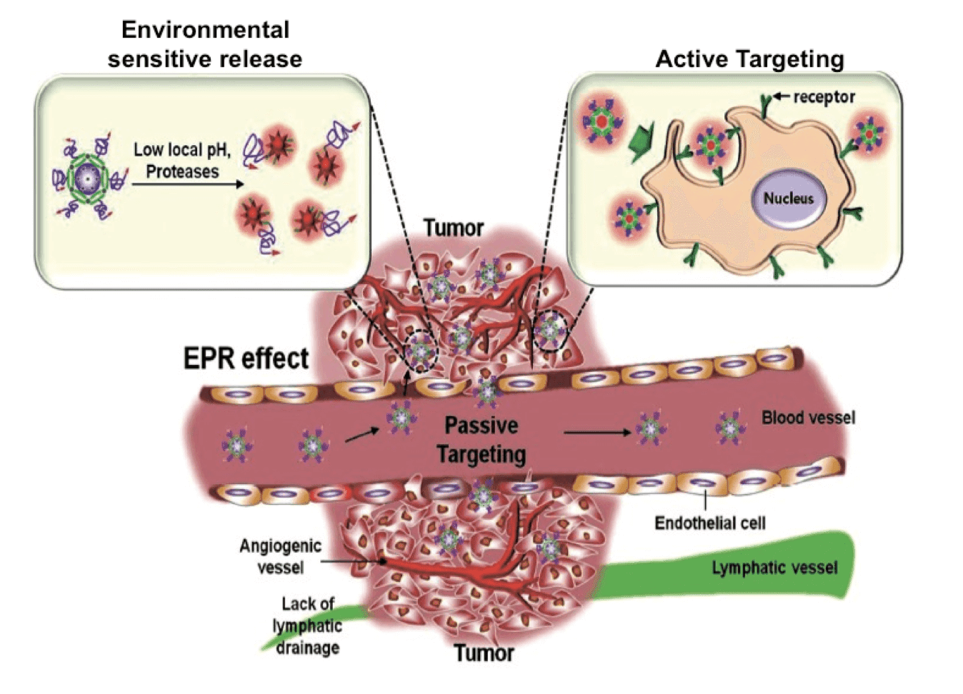
 Schematic illustration of the delivery of therapeutic agents to tumor tissues via EPR effect mediated passive targeting, the specific antibody-antigen recognition mediated active targeting of therapeutic agents to antigen/receptor-expressing tumor cells, and the environmental sensitive release of the payloads triggered by pH, redox pressure or proteases (Quant. Imaging Med. Surg., 2012).
With over a decade’s experiences in therapeutic antibody and ADC analysis, Creative Biolabs provide in vitro analysis of ADC targeting ability by using both 2D and 3D cell culture models. The 2D single cell type culture and mixed cell type co-culture models can provide primary indication of the ADC targeting ability for fast and high throughput analysis. While the 3D cell culture and micro-tissue models, presented by our 3D cell culture platform, at Creative Biolabs can provide precise assessment of the ADC targeting profile. In particular, with the expertise in 3D cell culture, our scientific team at Creative Biolabs also helps customers with the establishment of special 3D tumor micro-tissue models to fulfill the requirement of specific ADC analysis. Via comparative analysis using 2D and 3D cell culture models, experienced scientists at Creative Biolabs provide customers with practical advices on the optimization of ADC products in a timely and cost efficient manner. Please contact us for more information and a detailed quote.
Reference:
- Park, K. Polysaccharide-based near-infrared fluorescence nanoprobes for cancer diagnosis. Quant. Imaging Med. Surg. 2012, 2(2):106-113.
For Research Use Only. NOT FOR CLINICAL USE.

Online Inquiry
Welcome! For price inquiries, please feel free to contact us through the form on the left side. We will get back to you as soon as possible.
