As an incontestable global leading provider of antibody-related services, Creative Biolabs has accumulated years of experience and expertise in the development of antibodies for research, diagnostic, and therapeutic applications. Now, Creative Biolabs offers a full range of in vitro diagnostic (IVD) antibody development services and immunoassay development services to global clients.
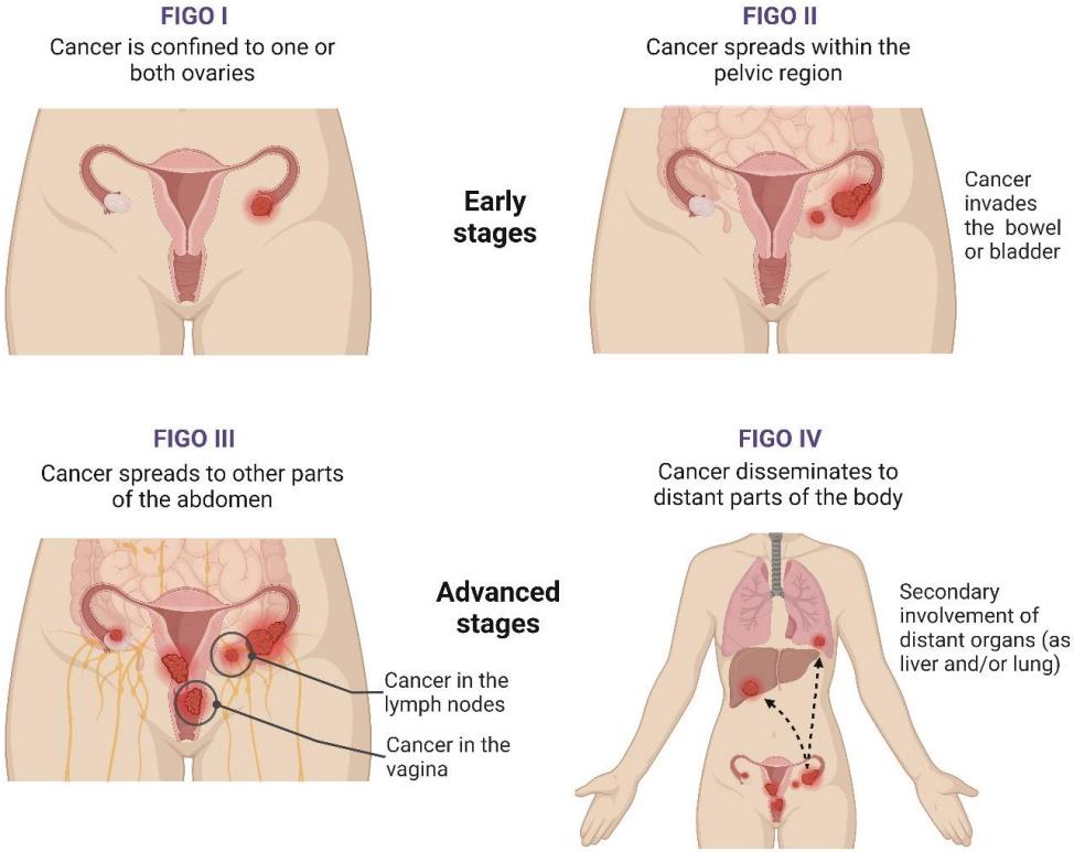
Ovarian cancer is the cancer that arises from the ovary. It causes more deaths among all the cancer types in the United States than any other type of female reproductive tract cancer, with approximately 20,000 new cases and 15,000 deaths in 2007. Ovarian cancer results in abnormal cells that invade or spread to other parts of the body. When this process starts, there may be no or only vague symptoms. Signs and symptoms become more and more noticeable as cancer progresses. These symptoms may include bloating, pelvic or abdominal pain, abdominal swelling with weight loss, and loss of appetite, trouble eating, fatigue, constipation. About 72% of ovarian cancers are diagnosed at advanced stage and approximately 30% women with ovarian cancers could survive 5 years.
 Fig.1 Stages of ovarian cancer.1
Risk Factors
Ovarian cancer is associated with the amount of time spent ovulating, which suggesting not having children is a risk factor for ovarian cancer. One explanation is that ovulation is suppressed via pregnancy. During ovulation, cells are constantly stimulated to divide while ovulatory cycles continue. Therefore, people who have not borne children are at higher risk of catching ovarian cancer than those who have. Meanwhile, a longer period of ovulation caused by early first menstruation and late menopause is also a risk factor. Both obesity and hormone replacement therapy also increases the risk.
Women who have fewer menstrual cycles, no menstrual cycles, breastfeeding have a lower risk of developing ovarian cancer. Also, the same can be said for the women who take oral contraceptives, have multiple pregnancies, and have a pregnancy at an early age. Women who have had the tubal ligation, both ovaries removed, or hysterectomy have a reduced risk of developing ovarian cancer. It is noteworthy that age is also a risk factor.
Type
Tumors of surface epithelial origin constitute about two-thirds of all ovarian neoplasms and the proportion of ovarian malignant neoplasms is probably even greater. They occur predominantly in adults, the malignant forms generally appear later in life. Epithelial tumors are classified into several subtypes according to the predominant pattern of differentiation of the tumor cells.
Serous neoplasms are one of the most common neoplasms. Serous cystadenoma is often a unilocular cyst, with a smooth surface and filled with serous fluid, and sometimes consists of multilocular cysts. Adenofibroma is generally a solid fibrous tumor. Serous carcinoma is usually large and bilateral exhibiting a mixture of cystic, papillary, and solid growth patterns. The carcinoma often invades the ovarian capsule and grows on the surface of the ovary. Foci of necrosis and hemorrhage are very common in serous carcinoma.
Mucinous cystadenoma is also one of the most common ovarian neoplasms. Macroscopically, mucinous tumors are approximately the largest of all ovarian tumors and is usually a cystic unilocular neoplasm with a thin, smooth wall and filled with watery mucinous fluid. Borderline and invasive mucinous tumors often have soft papillae and solid areas. Mucinous carcinomas are predominantly solid. Necrosis and hemorrhage are more common in invasive than borderline and benign mucinous tumors.
Endometrioid tumors are characterized by the presence of epithelial elements, stromal elements, or both of these two, which resemble those of the endometrium. Ovarian endometriosis has a range of characteristics, consistency and colors and thus is characterized by the presence of small blue, red and brown spots and patches associated with scars and dense fibrous adhesions. The content of cysts is often dark brown. Benign and borderline endometrioid tumors are very rare. An endometrioid cystadenofibroma is composed of glands lined by endometrioid-type cells within fibrous.
Management
Once ovarian cancer is determined, the treatment should be scheduled and developed. The treatment usually is about surgery and chemotherapy, and sometimes radiotherapy should also be involved, regardless of the ovarian cancer subtype. Surgical treatment is presumably sufficient for well-differentiated malignant tumors and confined to the ovary. Addition of chemotherapy may be required for more aggressive tumors confined to the ovary. For patients with advanced disease, a combination of surgical reduction with a combination chemotherapy regimen is an ideal option. Also, the surgical treatment is sufficient for treating borderline tumors, even following spread outside of the ovary, and chemotherapy does not work so well in this case.
Creative Biolabs offers turn-key or ala carte services customized to suit our client's needs. If you are interested in our platforms or our services, please contact us for more detail information.
Our services target the following potential biomarkers for ovarian cancer diagnosis:
| L1CAM | CD24 | ADAM10 | EMMPRIN | Claudin | miR-21 |
| miR-141 | miR-200a | miR-200b | miR-200c | miR-203 | miR-205 |
| miR-241 | CA125 | BRCA1 | BRCA2 | hMLH1 | hMSH2 |
| APC | p14ARF | p16INK4a | DAPKinase | CLDN3 | FOLR1 |
| COL18A1 | CCND1 | FLJ12988 | miR-214 | miR-199a | miR-140 |
| miR-125b1 | miR-92 | miR-93 | miR-126 | miR-29a | miR-155 |
| miR-127 | miR-99b | Mesothelin | HE4 | VEGF-C |
Reference
For Research Use Only.
