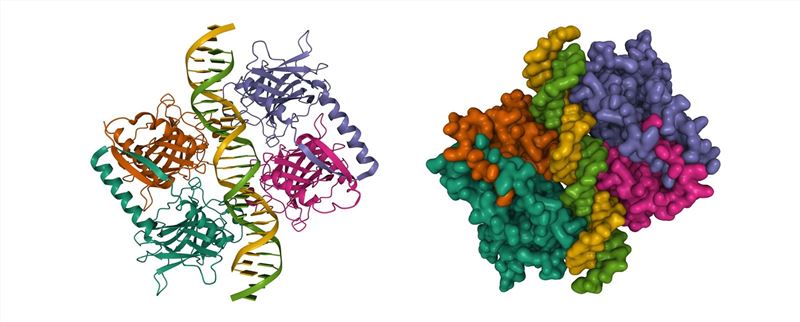
Proteins can undergo reactions such as thermal denaturation and unfolding when exposed to heat, which can result in denaturation and loss of protein activity. However, there is a growing demand for heat-stable proteins in various research and applications, particularly in the field of biomedicine. As the use of proteins continues to expand across different industries, the need for stable variants of protein domains is on the rise. Drawing upon our extensive expertise and phage display platform, Creative Biolabs provides comprehensive services to facilitate the development of thermal stable variants of protein domains.

The thermal stability of proteins plays a crucial role in both research and practical applications. In high-temperature research environments, heat-stable proteins exhibit a significant advantage by retaining their proper folding and activity. Even at room temperature, these proteins can maintain their folded state and functionality. Notably, thermal stable proteins have been found to accumulate in E. coli, primarily due to their propensity to adopt less aggregation-stable structures, resulting in high solubility even at elevated concentrations.
Phage display technology is a valuable method for screening stable variants within protein domains, such as the common thermal stable variants of protein domains and protease-stable variants of protein domains. Phage particles are essentially stable macromolecules, capable of maintaining their integrity and infectivity under conditions that could disrupt the three-dimensional structure or binding of displayed proteins or structural domains. For example, phage M13 particles can withstand exposure to conditions like 65°C heat, 4 M urea, 50 mM dithiothreitol, 5 M guanidinium chloride, highly acidic (i.e., pH 2) or alkaline (i.e., pH 12) solutions, various organic solvents, and proteases such as trypsin, chymotrypsin, endoproteinase Glu-C, and proteinase K, without losing infectivity. Therefore, if a wild-type protein structural domain loses its three-dimensional structure or binding capability under specific conditions, it is possible to generate mutagenic libraries and select for folded stable proteins. The preservation of the displayed protein structural domain's structure and function depends on the inherent thermal stability of the protein or structural domain itself, rather than being influenced by protein III or the viral particle. For example, a study utilized phage display technology to screen variants of the Bacillus subtilis endoxylanase (XynA) protein. The process involved three rounds of screening at progressively higher temperatures to obtain the desired thermal stable variants of protein domains.
Currently, there are three commonly used methods to determine the midpoint temperature (Tm) of thermal denaturation in proteins. They are differential scanning calorimetry, circular dichroism spectroscopy, and differential scanning fluorescence. Differential scanning calorimetry is a technique that measures heat absorption and release by comparing the power delivered to the object being measured with a reference object as a function of temperature under controlled temperature conditions. It quantitatively measures thermodynamic parameters and provides insights into thermal effects associated with conformational changes during protein denaturation. Circular dichroism spectroscopy analyzes structural characteristics of biological macromolecules, including proteins, nucleic acids, and saccharides. It utilizes polarized light passed through chiral structures to determine the circular dichroism, offering valuable information about secondary protein structures. Differential scanning fluorescence methods involve two approaches: one with added exogenous fluorescent dyes and one without. Both methods expose protein hydrophobic groups for detection. In the former, a fluorescent dye affinitive to the protein's hydrophobic region is added, and changes in fluorescent signals are tracked during warming to determine Tm. In the latter, Tm is measured by detecting changes in the hydrophobic environment of aromatic amino acids (tryptophan and tyrosine) during protein unfolding.
At Creative Biolabs, we possess extensive knowledge and experience in screening thermal stable variants of protein domains. Our team is ready to share our insights and experience with you.
All listed services and products are For Research Use Only. Do Not use in any diagnostic or therapeutic applications.
| USA:
Europe: Germany: |
|
|
Call us at: USA: UK: Germany: |
|
|
Fax:
|
|
| Email: info@creative-biolabs.com |
