Virus-mediated Labeling Exosomes Development Services
Exosomes are released outside the cell in the form of exocrine after the fusion of multivesicular bodies in the cell and cellular membrane. According to the mechanism of exosome generation, the modification of the exosome membrane can be achieved by genetic engineering of parental cells. The fluorescent protein is a commonly used reporter protein, which can be excited by excitation light of a specific wavelength to emit a corresponding fluorescent signal. Parental cells can be genetically modified by infection with viral vectors that insert fluorescent protein-coding sequences and exosomal membrane protein-coding sequences. These modified cells then secrete exosomes with fluorescent proteins, allowing the exosomes to be tracked from donor to recipient cells. Creative Biolabs can provide a series of viral vectors that can be directly used to infect parental cells, helping customers to observe the traces of exosomes.
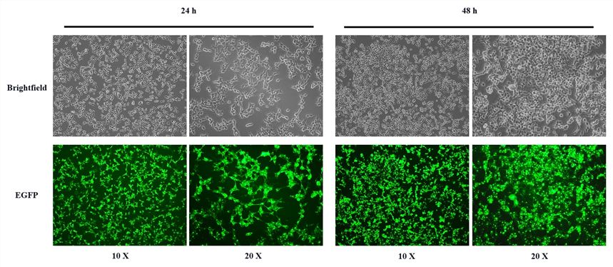
 Fig.1 Images of HEK293T cells transfected with the CD63-EGFP plasmid at 24- and 48-hours post-transfection.
Abundant Viral Vectors Available for Exosome Labeling
Replication-defective recombinant viruses, such as lentiviruses, in which virulence-related genes have been deleted, have been widely used in genetic engineering. This virus can effectively introduce and integrate exogenous genes into the genome of target cells through in vitro infection, to achieve long-term expression of exogenous genes in dividing and non-dividing cells. Lentiviruses can infect a wide range of host cells, including tumor cell lines, fibroblasts, neurons, lymphocytes, and macrophages. On exosomal membranes, the tetraspanin family (including CD63, CD81, and CD9) involved in exosome trafficking is highly expressed. Therefore, these exosomal membrane proteins and fluorescent proteins can be constructed into lentiviruses. These lentiviral vectors can be used to infect parental cells. Fluorescent membrane proteins are expressed on the exosome membrane, which can be used to track the formation, secretion, targeting, and trafficking mechanisms of exosomes.
Creative Biolabs Features
-
The lentiviral vectors developed by Creative Biolabs can express fusion proteins through the CMV promoter in most mammalian cells.
-
These lentiviral vectors have high biological safety, a wide range of hosts, and high lentiviral titers, which can reach 10^9 TU/mL.
-
Creative Biolabs provides lentiviral vectors and purified fluorescent exosomes carrying different fluorescent markers (including GFP, BFP, and mCherry), which can meet customers' requirements for color.
-
If you have innovative ideas and needs, Creative Biolabs can provide you with personalized fluorescent exosome preparation services.
 Fig.2 Fusion strategy of exosomal membrane proteins and fluorescent proteins.
Creative Biolabs is the leading provider of exosome services and products. We can provide one-stop services including exosome extraction, exosome identification, exosome modification, exosome profiling, and exosome function verification in vivo and in vitro. Please contact us to put forward your needs and ideas to provide you with the most suitable products, or formulate the most suitable solutions.
Reference
-
Levy, D.; Do, MA.; et al. Genetic labeling of extracellular vesicles for studying biogenesis and uptake in living mammalian cells. Methods in Enzymology. 2020. 645:1-14.
For Research Use Only. Cannot be used by patients.
Related Services:

 Fig.1 Images of HEK293T cells transfected with the CD63-EGFP plasmid at 24- and 48-hours post-transfection.
 Fig.2 Fusion strategy of exosomal membrane proteins and fluorescent proteins.


