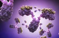
Creative Biolabs is offering the most comprehensive services for antibody development projects. With strict regulation and effective execution, we are dedicated to providing the most valuable solutions to complete your projects.
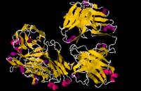
With over a decade of experience in phage display technology, Creative Biolabs can provide a series of antibody or peptide libraries that are available for licensing or direct screening. These ready-to-use libraries are invaluable resources for isolating target-specific binders for various research, diagnostic or therapeutic applications.
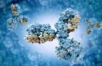
Creative Biolabs has established a broad range of platforms for developing novel antibodies or equivalents. These cutting-edge technologies enable our scientists to meet your demands from different aspects and tailor the most appropriate solution that contributes to the success of your projects.
With deep understanding in antibody-related realms and extensive project experience, Creative Biolabs offers a variety of references to help you learn more about our capacities and achievements, including infographic, flyer, case study, peer-reviewed publications, and all kinds of knowledge that can assist your projects. You are also welcome to contact us directly for more specific solutions.
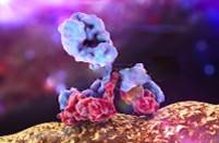
Get a real taste of Creative Biolabs, one of the most professional custom service providers in the world. We are committed to providing highly customized comprehensive solutions with the best quality to advance your projects.
This membrane protein is Human Akt1 (19-244). It has been tested in SDS-PAGE. We provide this protein to facilitate your membrane protein antibody discovery and development.
Product Specifications
Host Species
Human
Target Protein
Akt1
Protein Length
ECD
Molecular Weight
The protein has a calculated MW of 59.5 kDa. The protein migrates as 60-66 kDa under reducing (R) condition (SDS-PAGE) due to glycosylation.
Sequence
AA Met 1 - Ala 480 (Accession # AAH00479).
Product Description
Application
SDS-PAGE
Expression Systems
HEK293
Tag
His tag at the C-terminus, followed by a twin strep tag
Protein Format
Soluble
Reconstitution
Please see Certificate of Analysis for specific instructions.
Endotoxin
<1.0 EU/μg by the LAL method
Purity
>92% as determined by SDS-PAGE.
Buffer
Lyophilized from 0.22 μm filtered solution in 20 mM Tris, 150 mM NaCl, pH8.0. Normally trehalose is added as protectant before lyophilization.
Target
Target Protein
Akt1
Full Name
AKT serine/threonine kinase 1
Introduction
This gene encodes one of the three members of the human AKT serine-threonine protein kinase family which are often referred to as protein kinase B alpha, beta, and gamma. These highly similar AKT proteins all have an N-terminal pleckstrin homology domain, a serine/threonine-specific kinase domain and a C-terminal regulatory domain. These proteins are phosphorylated by phosphoinositide 3-kinase (PI3K). AKT/PI3K forms a key component of many signalling pathways that involve the binding of membrane-bound ligands such as receptor tyrosine kinases, G-protein coupled receptors, and integrin-linked kinase. These AKT proteins therefore regulate a wide variety of cellular functions including cell proliferation, survival, metabolism, and angiogenesis in both normal and malignant cells. AKT proteins are recruited to the cell membrane by phosphatidylinositol 3,4,5-trisphosphate (PIP3) after phosphorylation of phosphatidylinositol 4,5-bisphosphate (PIP2) by PI3K. Subsequent phosphorylation of both threonine residue 308 and serine residue 473 is required for full activation of the AKT1 protein encoded by this gene. Phosphorylation of additional residues also occurs, for example, in response to insulin growth factor-1 and epidermal growth factor. Protein phosphatases act as negative regulators of AKT proteins by dephosphorylating AKT or PIP3. The PI3K/AKT signalling pathway is crucial for tumor cell survival. Survival factors can suppress apoptosis in a transcription-independent manner by activating AKT1 which then phosphorylates and inactivates components of the apoptotic machinery. AKT proteins also participate in the mammalian target of rapamycin (mTOR) signalling pathway which controls the assembly of the eukaryotic translation initiation factor 4F (eIF4E) complex and this pathway, in addition to responding to extracellular signals from growth factors and cytokines, is disregulated in many cancers. Mutations in this gene are associated with multiple types of cancer and excessive tissue growth including Proteus syndrome and Cowden syndrome 6, and breast, colorectal, and ovarian cancers. Multiple alternatively spliced transcript variants have been found for this gene.
Alternative Names
AKT; PKB; RAC; PRKBA; PKB-ALPHA; RAC-ALPHA; RAC-alpha serine/threonine-protein kinase; AKT1m; PKB alpha; RAC-PK-alpha; protein kinase B alpha; proto-oncogene c-Akt; rac protein kinase alpha; serine-threonine protein kinase; v-akt murine thymoma viral oncogene homolog 1; v-akt murine thymoma viral oncogene-like protein 1