Cell therapy approaches are potential new therapies to repair damaged organs or induce stronger immunity killing function. Empowered by leading technologies as well as years of experience in this field, Creative Biolabs offers in vivo assay development services in animal model to meet every customer's needs.
In vivo assays for stem cell research refer to experiments conducted on live organisms to study the behavior, function, and therapeutic potential of stem cells. These assays are an essential part of stem cell research as they provide crucial insights into how stem cells interact with their environment, differentiate into specialized cell types, and contribute to tissue regeneration and repair.
In vivo assays involve introducing stem cells into an animal model, such as mice or rats, and monitoring their behavior and impact on the host organism. These experiments help researchers understand the factors that influence stem cell proliferation, differentiation, and integration into tissues, as well as assess the safety and efficacy of potential stem cell-based therapies.
Some common in vivo assays for stem cell research include transplantation studies, where stem cells are transplanted into a specific tissue or organ to investigate their regenerative potential, as well as lineage tracing studies, where stem cells are labeled to track their fate and differentiation in vivo.
At Creative Biolabs, we provide customized in vivo assay development service based on your purpose. Our capabilities include but not limited to:
We provide various histological section preparation and analysis including paraffin section, frozen section and ultrathin section. We also provide immunohistochemistry service.
Non-invasive imaging technologies have been developed to replace the traditional invasive histological analysis. Our tracking imaging service for cells tracking include live imaging of small animals, magnetic resonance imaging (MRI), positron emission tomography (PET) and single-photo emission computed tomography (SPECT).
Direct labeling is the most straightforward and widely used method, and is accomplished by introducing a marker into the cells or onto the cell surface before implantation. We provide diversified cell labeling service including fluorescent probe direct labeling, magnetic labeling, and radionuclide direct labeling.
At Creative Biolabs. our experienced scientists and specialized technology ensure reasonable project design to achieve an adequate in vivo assay. We have various experimental technology platforms for you to choose. Please don't hesitate to contact us for more information.
Below are the findings presented in the article related to the in vivo assay for stem cells.
Stem cell therapy has great potential for a variety of regenerative medicine applications. However, clinical stem cell therapies are severely limited by the challenge of assessing the location and functional status of implanted cells in vivo. Anamik Jhunjhunwala et al. presented a multidisciplinary approach for spatial tracking and functional assessment of transplanted stem cell fates using caspase-3 responsive nanosensors.
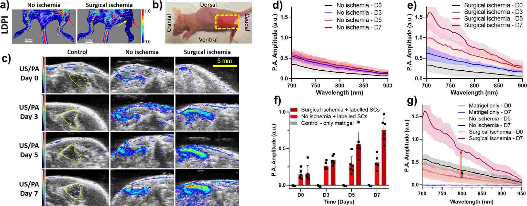
They sought to validate the AuPD nanosensor in a preclinical US/PA imaging scenario by demonstrating spatial tracking of injected stem cells and apoptosis monitoring in an animal model. The results showed that their nanosensor could be used for longitudinal and non-invasive tracking and apoptosis monitoring of injected stem cells, as demonstrated in an in vivo apoptosis model in mice.
 Fig. 1 Characterization of the in vivo activity of the AuPD nanosensor.1
Reference
For Research Use Only. Not For Clinical Use.