Golgi Apparatus Positioning Intrabody Development Service
With the sophisticated searching and screening of the Golgi-retention peptide libraries, Creative Biolabs offers the top motifs which have survived in our strict validations. In terms of our novel Super™ Intrabody Development Platform, our scientists can tailor the best-fit strategy to develop special Golgi-positioning intrabodies for your research.
Golgi Apparatus Positioning
Golgi apparatus is a specific organelle found in most eukaryotic cells as one of the first organelles identified and observed in detail by the Italian scientist Camillo Golgi in 1897 and named after him in 1898. A single Golgi apparatus is usually located near the cell nucleus by the site of the centrosome, meanwhile, the multiple Golgi apparatus are stacked together by microtubules in mammals. On the spatial structure, Golgi apparatus maintains two networks primarily:
- The cis-Golgi network (CGN) is manifested as a collection of fused and flattened membrane-enclosed disks (cisternae), which is originated from vesicular clusters budded off the ER and maintaining versatile enzymes catalyzing early protein modifications.
- The trans-Golgi network (TGN) is the final cisternal structure, where mature proteins are packaged into vesicles destined to lysosomes, secretory vesicles, or the cell surface.
Golgi performs multiple vital bioprocesses as a part of the cellular endomembrane system. For instance, Golgi modifies proteins transported from ER post-translationally with the common glycosylation and phosphorylation, and other minor modifications, which may be phosphorylation of oligosaccharides on lysosomal proteins, the removal of mannose residues, addition of specific molecules (N-acetylglucosamine, galactose, sialic acid, etc.), sulfation of tyrosines and carbohydrates, and so on. Furthermore, Golgi is also a crucial site to synthesize proteoglycans via appending proteins to glycosaminoglycans and form a signal sequence by adding a mannose-6-phosphate label on proteins destined for lysosomes. Despite the depiction above, the overall function of Golgi manifests as a particular protein processing organelle to package mature proteins into membrane-bound vesicles inside the cell for secretion or destined subcellular structures. For instance, the antibody-secreting plasma B cells of the immune system have prominent Golgi complexes to process mature antibody and secret out of the cells. In addition, Golgi also involves in lipid transportation and lysosome formation, or other sophisticated bioprocesses remaining undiscovered.
Protein retention in the Golgi
Since the Golgi apparatus performs various unique roles, they maintain a population of resident proteins including multiple categories of the glycosyltransferases and glycosidases. Noticeably, the Golgi adopts multiple protein retention strategies than the simple retention signal peptides in other organelles. Currently, the scientific community summarized these strategies as the following:
- Protein aggregation/Kin-recognition: the formation of homo- or hetero-oligomers or protein aggregation is adopted for excluding retention proteins from transport vesicles.
- The transmembrane domain-mediated Golgi retention: the shorter transmembrane domains of Golgi retention proteins have more tendencies for the thinner Golgi membrane bilayer than the plasma membrane, meanwhile, the enrichment of amino acids with aromatic side chains in Golgi-resident SNARE proteins also prefers the particular ratio of lipids (glycerophospholipids to sphingolipids) of the Golgi membrane. Thus, the type I and type II proteins can be reliably predicted from the length and amino acid composition of the transmembrane for specific intracellular localization.
- The cytoplasmic tail: functions to anchor Golgi-resident proteins via stable interaction with cytoplasmic chaperones or other proteins specifically localized on the Golgi outer membrane.
- The transmembrane luminal stem region: functions similarly as the cytoplasmic tail to anchor Golgi-resident proteins.
- The signal peptide-based Golgi retention: a few Golgi-resident proteins possess a signal peptide-based motif for their steady-state localization, which relies on a dynamic process in which proteins are retained at steady-state via successive recycle of retrograde and anterograde transport.
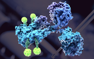
 Fig.1 Intrabody scFv (PDB: 5B3N) targeting to the Golgi apparatus.
Other Anchoring Modification Options provided by Creative Biolabs
Including the Golgi apparatus, Creative Biolabs can also develop specific positioning antibody for a variety of destination, which including but not limited to:
| Extracellular Space | Extracellular Plasma Membrane | Intracellular Plasma Membrane | Cytoplasm |
| Nucleus | Endoplasmic Reticulum | Golgi Apparatus | Mitochondria |
| Chloroplast | Lysosome | Peroxisome | Vacuoles |
Since our platform possesses the most professional technology and accumulative experience, Creative Biolabs is confident in offering the best intrabody development service to our clients all over the world. Please inquire us for detailed information.
All of our antibody products and services can only be used for preclinical research studies. Do not use them on humans.
Related Services:
- Extracellular Space Positioning Antibody Development
- Extracellular Plasma Membrane Positioning Antibody Development
- Intracellular Plasma Membrane-Anchored Intrabody Development
- Cytoplasm Positioning Intrabody Development
- Nucleus Positioning Intrabody Development
- Endoplasmic Reticulum Positioning Intrabody Development
- Mitochondria Positioning Intrabody Development
- Chloroplast Positioning Intrabody Development
- Lysosome Positioning Intrabody Development
- Peroxisome Positioning Intrabody Development
- Vacuole Positioning Intrabody Development

Welcome! For price inquiries, please feel free to contact us through the form on the left side. We will get back to you as soon as possible.
Contact us
USA
Tel:
Fax:
Email:
UK
Tel:
Email:
Germany
Tel:
Email:


