Creative Biolabs is pleased to introduce our iPSC-facilitated melanin content quantification service to our global customers, to advance your projects on pigmentation and AMD.
Melanin is a type of biological pigment recognized as an amorphous polymer, produced and stored in melanocytes, a group of cells in the basal layer of the epidermis. In humans, melanin is primarily distributed in skin, mucous membranes, hair, retina, pia mater, and other tissues. There are 5 basal types of melanin, among which eumelanin, pheomelanin, and neuromelanin are common and play various roles in physiological processes and diseases in humans.
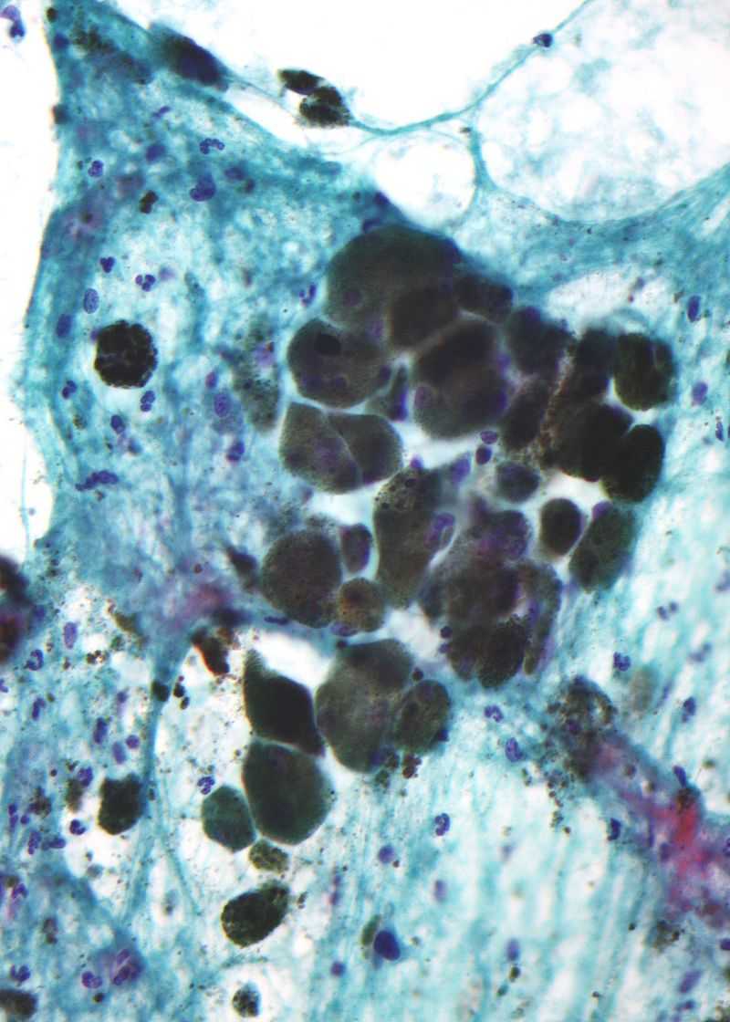 Fig.1 Micrograph of melanin pigment in a pigmented melanoma.1
Melanin is involved in the regulation of cellular metabolism, generating an oxidative environment. It exhibits a variety of unique physicochemical properties, including optical absorption, electrical conductivity, and affinity to a wide range of organic/inorganic compounds.
Possessing radioprotective and scavenging properties, melanin induces pigmentation and affects the behavior of melanoma cells. Also, melanin is the major pigment to absorb light in retinal pigment epithelium (RPE) and its loss in RPE indicates ocular senescence, serving as a risk factor and a signature symptom of age-related macular degeneration (AMD). Melanin content quantification is a tool that cannot be ignored to study the pathological role of RPE in ocular senescence and the occurrence and progression of AMD. In addition, quantification of melanin can provide important information for differentiating melanoma from benign pigmented lesions and in assessing pigmentary diseases.
There are various non-invasive methods of melanin content quantification available now, including elemental analysis, MALDI-MS, HPLC, Pump-probe, Raman, EPR, NMR, FTIR, UV/visible spectroscopy, pyrolysis, etc.
Depending on classifying melanin pigments based on their melanin structural units, elemental analysis, Py-GC–MS, ToF-SIMS, Raman, and MALDI are excellent choices if only qualitative information is required. If you want to further clarify the macromolecular structure, microscopy techniques (TEM and SEM), MALDI, Raman, FTIR, and NMR are suitable methods. Additionally, basic measurements (e.g. colorimetry, solubility, and UV/visible spectra), are also cost-effective and rapid approaches to measure melanin content. You can choose the most appropriate service according to your research requirements.
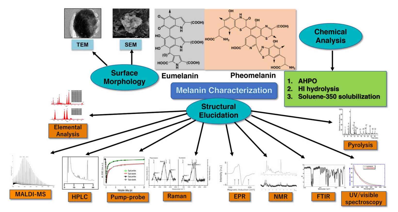 Fig.2 Various methods of melanin content quantification.1
Creative Biolabs is devoted to providing iPSC-facilitated melanin content quantification services and related customized services for our global clients to assist in your projects on pigmentation. With novel technical platforms and a professional team of scientists, we are pleased to offer our state-of-art products and services. If you want to learn more about detailed information, please feel free to contact us at any time.
References
For Research Use Only. Not For Clinical Use.