Creative Biolabs is pleased to introduce our iPSC-facilitated MITF expression assay service to our global customers, to advance your projects on pigmentation.
MITF (Microphthalmia-associated transcription factor, also called CMM8, bHLHe32) is a b-HLH-Zip transcription factor, which is related to lineage-specific pathway regulation of a variety of cells including mast cells, osteoclasts, and melanocytes. Several isoforms of MITF are produced by MITF gene because of alternative first coding exons in the promoter, including MITF-A/B/C/D/E/H/J/ Mc/M.
MITF participates in multiple signaling pathways including MAPK/BRAF/ERK, GSK-3, receptor tyrosine kinase KIT, mTOR, PI3K, SRC, AKT, P38, etc., and there are various MITF regulators, often modified in melanoma, up/down-regulating the expression level of MITF.
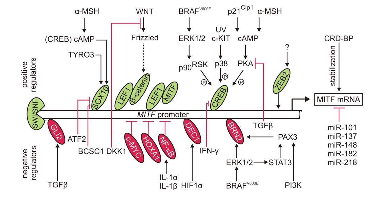 Fig.1 Transcriptional and post-transcriptional regulation of MITF expression.1
MITF is the primary modulator of melanocyte cell identity, controlling the production of melanin responsible for the colors of skin, hair, and eyes, and serves as a key regulator in skin pigmentation mechanisms. MITF plays essential roles in the production of pigment, as well as the differentiation, proliferation, and survival of melanocytes. Accordingly, deficiency of MITF expression in human embryonic stem cell differentiation leads to disorder in retinal pigment epithelium development and optic vesicle cell proliferation. Meanwhile, there are also feedback mechanisms between cellular activities and factors influencing the expression and activity of MITF, revealing the complexity of the regulation network.
Regarding the critical role of MITF expression in pigmentation, MITF expression assay is a highly frequently used tool in studies on pigmentation and other research related to melanocytes.
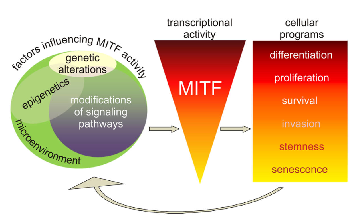 Fig.2 MITF expression and activity in melanoma cells.1
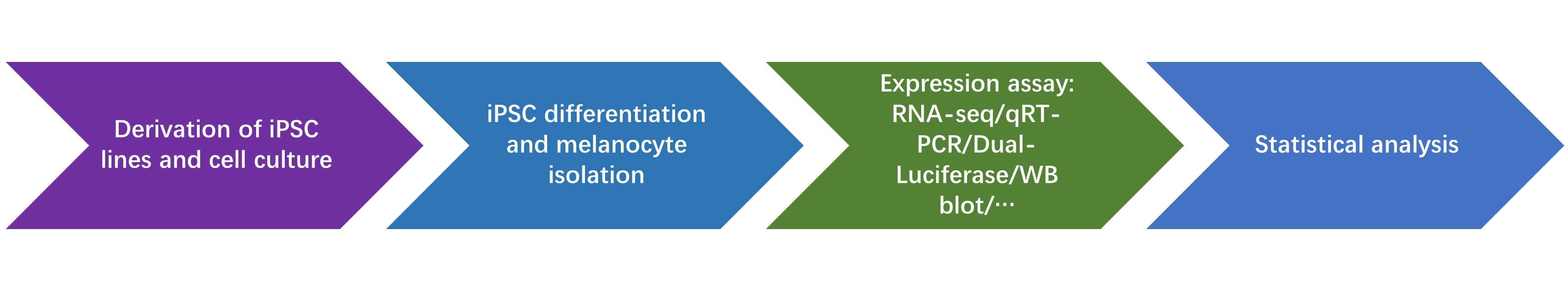
Induced pluripotent stem cells (iPSCs) are ideal sources of melanocytes and are valuable in studies on pigmentation for clinical, research, or commercial purposes. At Creative Biolabs, we provide iPSC-facilitated MITF expression assay service here with various alternative methods to analyze the expression of MITF, including but not limited to RNA-seq, qRT-PCR, Dual-luciferase, WB blot, and other experiments. We offer meticulous and customized experiment design, standardized operation, reliable data analysis, and detailed reports, to assist you in your exploration of the melanogenesis pathway.
Creative Biolabs is devoted to providing iPSC-facilitated MITF Expression Assay services and related customized services for our global clients to assist in your projects on pigmentation. With novel technical platforms and a professional team of scientists, we are pleased to offer our state-of-art products and services. If you need more detailed inquiries, please do not hesitate to contact us at any time.
Reference
For Research Use Only. Not For Clinical Use.