The ability of antigen-specific lymphocytes to differentiate and proliferate into effector cells is a key characteristic of the immune response. Therefore, it is very important to detect the cell activity and function in vivo or in vitro. Scientists have sought for an efficient procedure with minimal disruption to monitor an immune response for a long time. Until now, the intracellular staining with carboxyfluorescein succinimidyl ester (CFSE) has become a common method to follow lymphocyte differentiation and proliferation in labs.
Based on its ability to penetrate the cell membrane, CFSE is used to monitor cell proliferation and estimate the times of cell division. Conjugated antibodies can be used to probe the change of surface marker or internal molecular expression. CFSE can easily penetrate the cell membrane and covalently bind to the protein in the process of cell division and proliferation. The fluorescence will be evenly distributed to the two offspring cells. And its intensity is half of the parent cells, according to this feature. Hence, the fluorescence intensity gradually reduced with the division of cells. In addition, the labeled cells can be observed for weeks in vivo so that they are normally used in the experiments of cells test and cells activity observation using fluorescent electron microscopy or confocal.
In order to measure the biochemical and biophysical features, stained cells should be analyzed and screened through flow cytometer assay that can rapidly, accurately and specifically analyze single cell in molecular level.
Creative Biolabs has developed a highly efficient ECIA™ CFSE T cell proliferation assay that combines cell proliferation assay and flow cytometry testing to accurately monitor the proliferation of CD4+ cells and the details of T cell responses. ECIA™ CFSE T cell proliferation assay can be applied to not only the preclinical screening of novel pharmaceutical proteins but also detection of the potential T cell epitopes. After communicating about the project objective with each customer. Creative Biolabs will design a cost and time efficient protocol and provide you a personalized service
Other optional ECIA™ cellular analysis services:
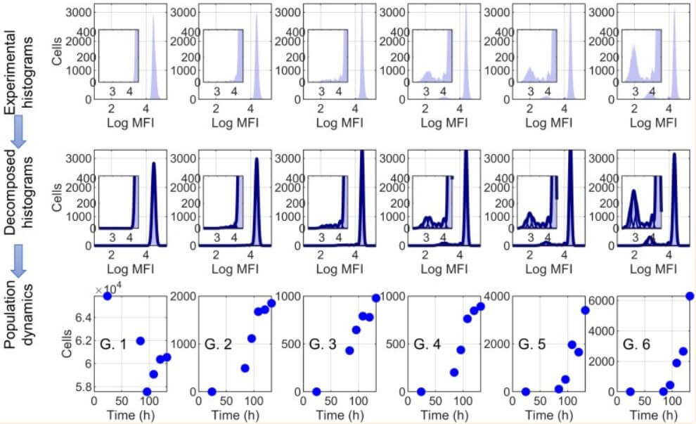
 Fig. 1 Mathematical analyses of CFSE-based proliferation assays. (Valerya Zheltkova, 2019)
The article focuses on the effects of PD-L1 inhibition in HIV-infected individuals, particularly examining how this treatment modality influences HIV-specific CD8 T cell responses. It highlights that PD-L1 blockade can significantly enhance the proliferative capacity of these T cells, which are crucial for controlling HIV. The study utilized the CFSE T cell proliferation assay to measure the proliferation of HIV-1 Gag-specific CD8 T cells from HIV-infected patients. This assay enabled the researchers to track the division of individual T cells and quantify the enhancement in T cell proliferation following PD-L1 blockade. The results from this method were pivotal in determining the dynamic parameters for HIV-specific T cells, which could then be modeled to predict potential clinical outcomes based on the PD-L1 blockade, revealing that the blockade might offer substantial benefits in managing HIV, particularly in slowing the disease's progression.
Carboxyfluorescein succinimidyl ester (CFSE) is a fluorescent dye used in cell proliferation studies. When cells are labeled with CFSE and then divide, the dye is halved between daughter cells, allowing researchers to track the number of cell divisions. This makes CFSE particularly useful for analyzing the proliferation of CD4+ T cells after stimulation with antigens or mitogens.
CFSE allows for the detailed tracking of cell division over multiple generations, which is crucial for understanding the dynamics of CD4+ T cell responses in immunological studies. By measuring the dilution of CFSE fluorescence, researchers can quantify the proliferation rate of CD4+ T cells in response to specific stimuli or under different experimental conditions.
The CFSE assay provides insights into the proliferation capacity of CD4+ T cells, which is a key aspect of their functionality. By analyzing how these cells expand in response to antigens, researchers can infer their health, activation status, and potential to provide help to other immune cells, which is crucial for effective immune responses.
The CFSE T cell proliferation assay involves several key steps: isolating T cells, labeling them with CFSE, stimulating them with antigens or mitogens, incubating for a specific period to allow cell division, and finally analyzing the cells using flow cytometry to measure the fluorescence intensity, which indicates the number of divisions the cells have undergone.
CFSE is an effective tool for studying T cell exhaustion by enabling the examination of proliferative responses under chronic stimulation conditions, such as those found in chronic infections or cancer. Researchers can assess how exhaustion affects the ability of CD4+ T cells to proliferate in response to their cognate antigens.
CFSE directly tracks cell divisions through dye dilution, providing a dynamic view of cell proliferation, whereas Ki-67 is a nuclear protein expressed in actively dividing cells but not in resting cells. Ki-67 staining requires cell fixation and permeabilization and only indicates whether a cell has been in the cell cycle recently, not the number of divisions.
CFSE itself does not differentiate between types of T cell responses but can be combined with other cell markers in multicolor flow cytometry assays to analyze different subsets of T cells, such as Th1, Th2, or regulatory T cells, based on their proliferation in response to specific conditions.
Use the resources in our library to help you understand your options and make critical decisions for your study.
All listed services and products are For Research Use Only. Do Not use in any diagnostic or therapeutic applications.
| USA:
Europe: Germany: |
|
|
Call us at: USA: UK: Germany: |
|
|
Fax:
|
|
| Email: info@creative-biolabs.com |
