Creative Biolabs presents IL-6 monitoring assays within an iPSC-derived sebocytes model to expedite the investigation of treatments for acne and hyper/hyposeborrhea.
Defined by an inflammatory etiology, acne stands as the most prevalent dermatological condition globally. Deviations in sebum secretion, abnormal keratinocyte proliferation, and dysbiosis in the microbiota, characterized by an imbalance in the population structure of Cutibacterium acnes, collectively contribute to the inflammatory state of acne-affected skin. The preservation of skin equilibrium is presumably dependent on a negative modulator of Nuclear Factor kappa B (NF-κB). Upon detection of C. acnes Pathogen-Associated Molecular Patterns (PAMPs) through Toll-like Receptor 2 (TLR-2) and TLR-4, NF-κB is activated, triggering the release of pro-inflammatory cytokines. Among the cytokines pivotal in orchestrating the immune response in acne is IL-6.
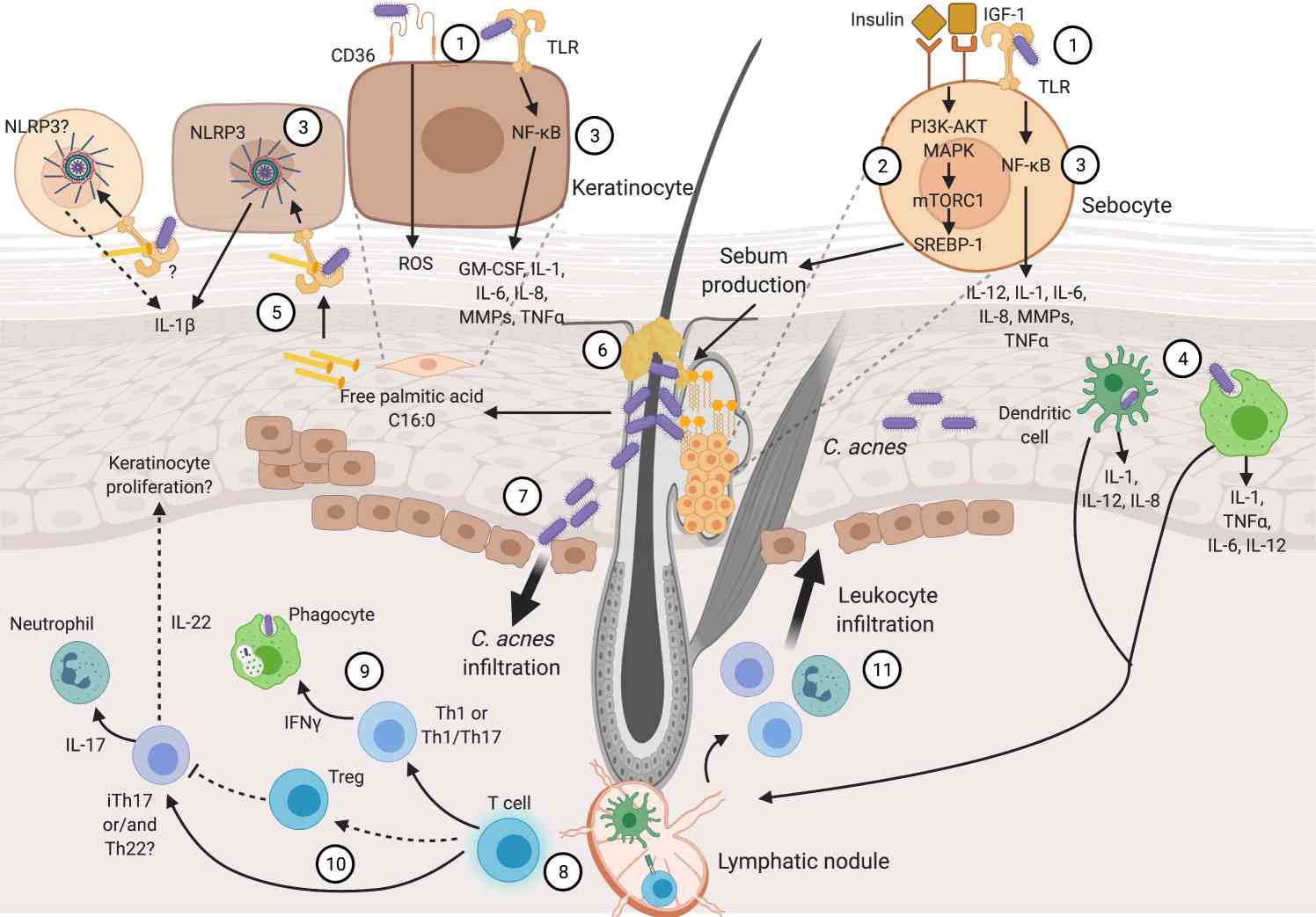 Fig.1 Inflammation mechanisms in acne pathophysiology.1
In patients prone to scarring, the immune response is sustained by T cells, neutrophils, and macrophages, along with B cell infiltration. Additionally, sebaceous glands may experience irreversible damage. In relation to T helper (Th) cells, increased concentrations of Th17/Th1 cells have been noted in atypical scar formation, coinciding with the deterioration of elastic and collagen fibers and diminished proliferation of epidermal cells. These phenomena are probably modulated by aberrant TGF-β1 signaling, which, in conjunction with IL-6, induces Th17 differentiation and subsequent secretion of pro-inflammatory IL-17. Consequently, activation of Th cells results in infiltration of leukocytes into the epidermis, activation of neutrophils by Th17 cells, and stimulation of phagocytes by Th1 or Th1/Th17 cells. Additionally, iTh17 cells and/or Th22 cells produce IL-22, potentially stimulating hyperkeratosis. These mechanisms contribute to the inflammatory responses evident in the affected dermis of individuals with acne.
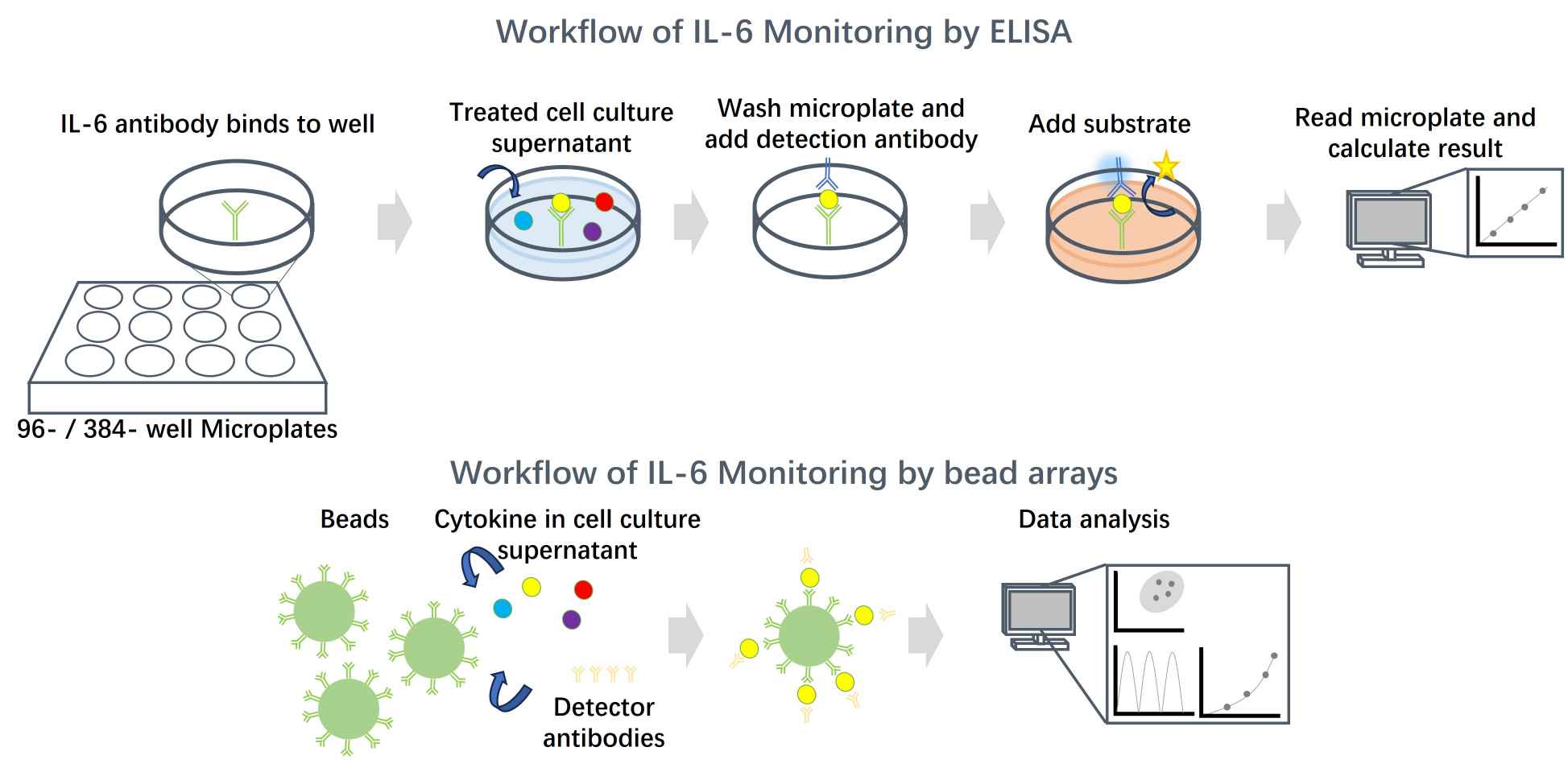
Creative Biolabs provides IL-6 monitoring assays from multiple angles. IL-6 secretion is examined using ELISA analysis, and bead arrays of cell culture supernatant. Additionally, a luciferase reporter system assay is utilized to assess the effectiveness of candidate acne drugs on the IL-6 promoter in iPSC-derived sebocytes.
To pioneer new avenues in scientific exploration within the fields of acne and hyperseborrhea, particularly in the evaluation and discovery of active pharmaceutical compounds, Creative Biolabs introduces a groundbreaking human sebocyte model derived from iPSCs. Renowned for their enhanced genomic stability, abundant availability, and exceptional reproducibility across batches, iPSC-derived cells stand as a robust resource for investigating treatments for Acne & Hyper/Hyposeborrhea. For more information or customized services, feel free to contact us.
Reference
For Research Use Only. Not For Clinical Use.