After extensive optimization research, Creative Biolabs provides highly reproducible and reliable sebocyte maturation assay based on induced pluripotent stem cells (iPSC)-derived sebocytes to assist you with drug development, screening, and evaluation targeting acne and hyper/hyposeborrhea.
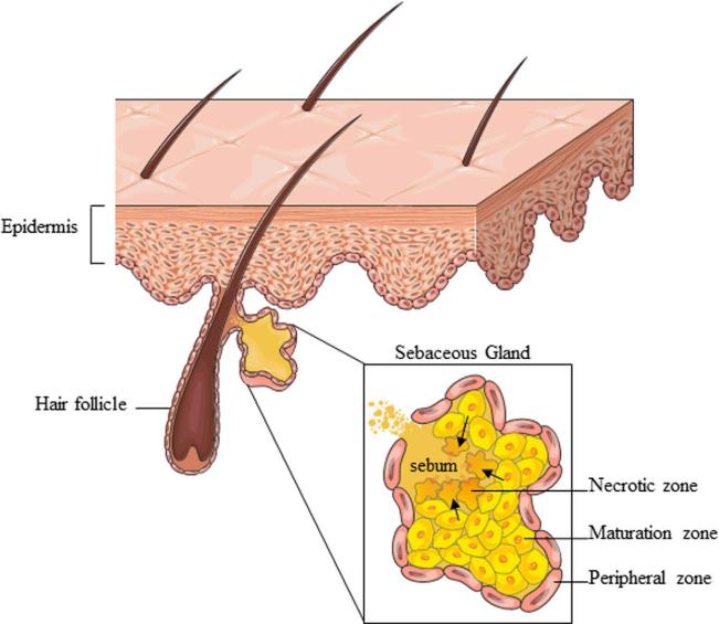
Sebocytes are specialized cells found in the sebaceous glands (SGs) of the skin responsible for sebum production. Derived from basal cells, sebocytes undergo a maturation process involving progressive lipid accumulation and differentiation into mature sebocytes. This process is crucial for the synthesis and secretion of sebum, an oily substance that lubricates and waterproofs the skin. During maturation, they progressively migrate toward the SG center and express markers. Mucin-1 (MUC1), a well-known marker of late differentiated sebocytes, interacts with growth factors and signaling pathways involved in regulating sebocyte differentiation and lipid production. Its expression levels have been linked to the maturation and functionality of sebocytes, highlighting its importance in sebum synthesis and skin homeostasis.
 Fig.1 The overall SG structure and partition into zones corresponding to various stages of sebocyte differentiation.1
Sebocyte maturation assay based on iPSC-derived sebocytes provides a robust model for investigating factors that influence sebocyte development, as well as for screening potential compounds aimed at modulating sebocyte maturation and regulating sebum production. In this assay, iPSCs are differentiated into sebocytes, mimicking the natural maturation process that occurs in the SGs. The key steps for sebocyte maturation assay focusing on the expression of the marker MUC1 are as follows.
1) Differentiation of iPSCs into Sebocytes: Induce the differentiation of iPSCs into sebocytes using appropriate growth factors and culture conditions that mimic sebaceous gland development.
2) Sebocyte Maturation: Allow the iPSC-derived sebocytes to mature in culture to resemble mature sebocytes found in the skin, ensuring they exhibit lipid accumulation and express sebaceous gland markers.
3) Treatment with Drug Candidates: Expose the mature iPSC-derived sebocytes to potential drug candidates or compounds of interest.
4) Immunofluorescence Staining: Fix the sebocytes and perform immunofluorescence staining to detect the expression of MUC1. Use specific antibodies against MUC1 for accurate detection.
5) Quantification and Imaging: Capture fluorescent images of the stained sebocytes using a fluorescence microscope. Quantify the intensity and distribution of MUC1 expression in response to the drug candidates.
6) Data Analysis: Analyze the results to assess the impact of the drug candidates on MUC1 expression and sebocyte maturation. Compare the expression levels with untreated control samples to evaluate the efficacy of the drug candidates.
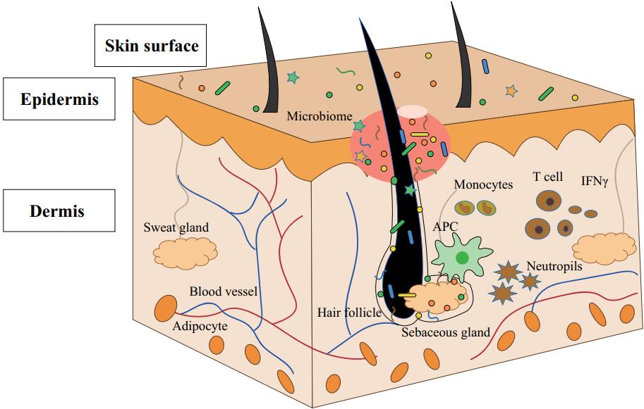
Dysregulated sebocyte maturation can lead to increased sebum production, contributing to acne pathogenesis. Hyperseborrhea is associated with acne, while hyposeborrhea may lead to skin dryness and potential barrier dysfunction, impacting acne susceptibility. Sebocyte maturation assay based on an iPSC-derived sebocyte model enables researchers to mimic the complex processes of sebocyte maturation and understand the mechanisms of sebocyte maturation, facilitating the discovery of new drugs for acne and hyper/hyposeborrhea. Additionally, this assay provides a platform for screening and evaluating potential drug candidates, ultimately facilitating the identification of compounds that could modulate sebocyte function, sebum production, and related pathways.
 Fig.2 Skin structure and pathogenesis of acne.2
With a professional team and rich experience, Creative Biolabs provides a full range of high-quality assay services to our valued customers. Please contact us for more information on iPSC services.
Reference
For Research Use Only. Not For Clinical Use.