Creative Biolabs is offering the most comprehensive services for antibody development projects. With strict regulation and effective execution, we are dedicated to providing the most valuable solutions to complete your projects.
HighlightsCreative Biolabs is offering the most comprehensive services for antibody development projects. With strict regulation and effective execution, we are dedicated to providing the most valuable solutions to complete your projects.
HighlightsCreative Biolabs is offering the most comprehensive services for antibody development projects. With strict regulation and effective execution, we are dedicated to providing the most valuable solutions to complete your projects.
HighlightsCreative Biolabs is offering the most comprehensive services for antibody development projects. With strict regulation and effective execution, we are dedicated to providing the most valuable solutions to complete your projects.
HighlightsWith over a decade of experience in phage display technology, Creative Biolabs can provide a series of antibody or peptide libraries that are available for licensing or direct screening. These ready-to-use libraries are invaluable resources for isolating target-specific binders for various research, diagnostic or therapeutic applications.
HighlightsCreative Biolabs has established a broad range of platforms for developing novel antibodies or equivalents. These cutting-edge technologies enable our scientists to meet your demands from different aspects and tailor the most appropriate solution that contributes to the success of your projects.
HighlightsWith deep understanding in antibody-related realms and extensive project experience, Creative Biolabs offers a variety of references to help you learn more about our capacities and achievements, including infographic, flyer, case study, peer-reviewed publications, and all kinds of knowledge that can assist your projects. You are also welcome to contact us directly for more specific solutions.
HighlightsGet a real taste of Creative Biolabs, one of the most professional custom service providers in the world. We are committed to providing highly customized comprehensive solutions with the best quality to advance your projects.
HighlightsImmune antibody libraries contain unique sets of antibody genes which originate from B cells taken from donors who have been immunized or who have experienced natural infection. Antibody libraries provide pre-optimized antibodies with high affinity and specificity against specific antigens because of previous immune exposure. The development of therapeutic antibodies from immune libraries eliminates the necessity for in vivo animal immunization.
The phage display method enables antibody fragments to be displayed on bacteriophage surfaces by genetically linking them to coat proteins such as pIII or pVIII found in M13 filamentous phages. The system facilitates fast identification and selection of antigen-specific antibodies by conducting multiple rounds of biopanning.
The fusion mechanism works as follows:
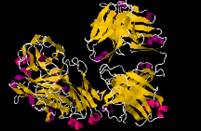
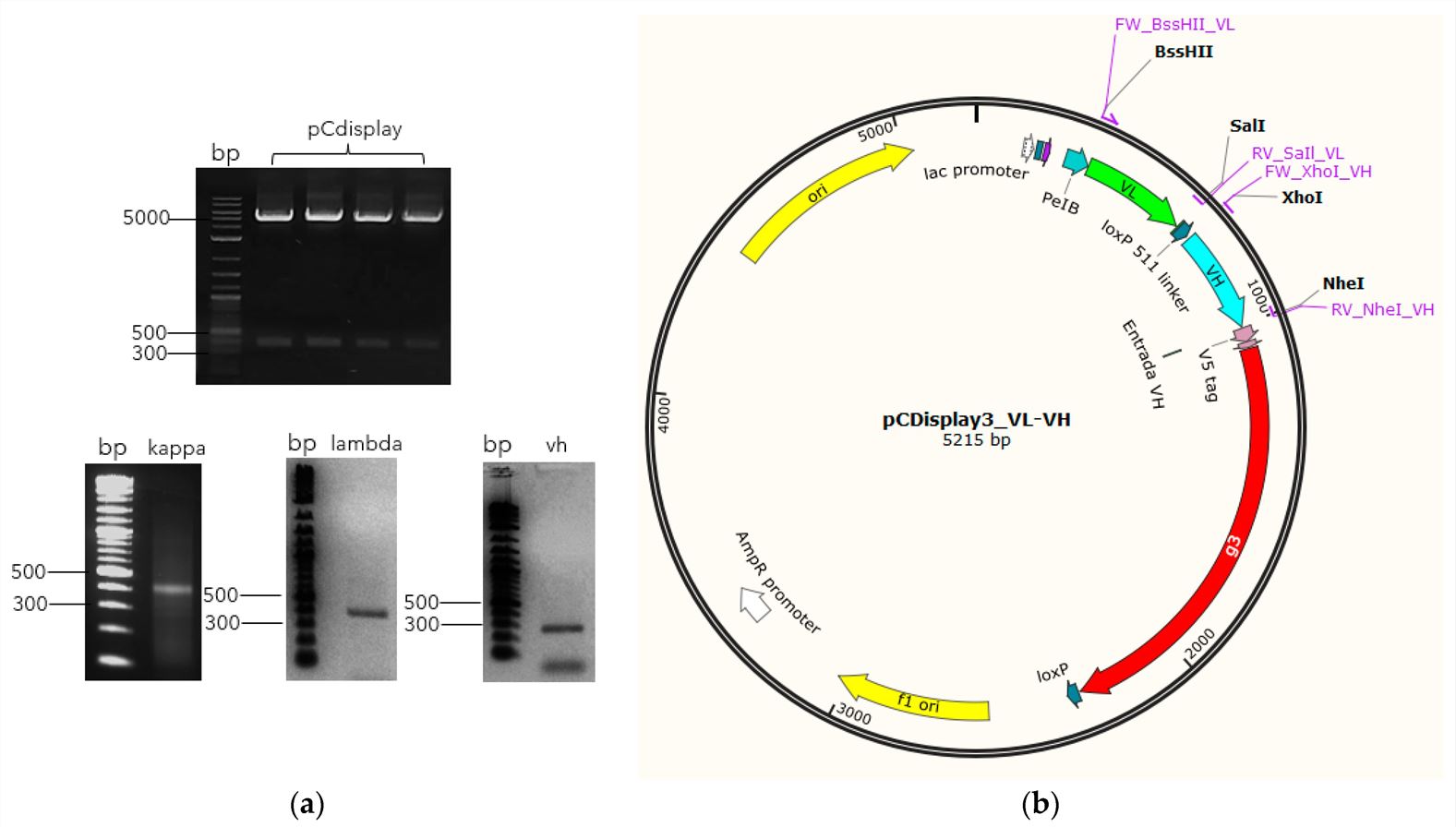 Fig. 1 Library assembly schematic.1, 3
Immune libraries are typically constructed from B cell-derived mRNA isolated from immune donors. The two major approaches are:
Key steps include:
Immune libraries are classified based on the source of antibody genes:
| Library Type | Description | Applications |
| Human Immune Library | Derived from human donors post-infection or vaccination | Therapeutic antibody discovery, clinical diagnostics |
| Humanized Library | Genetically modified to replace animal-derived framework regions with human sequences | Reduced immunogenicity in human therapy |
| Animal-Derived Library | Constructed from immunized animals such as mice, rabbits, or camels | Used in preclinical research, cross-species antibody generation |
| Feature | Immune Library | Naïve Library | Synthetic Library |
| Affinity | High, due to immune selection | Low, requires in vitro maturation | Variable, depends on design |
| Diversity | Moderate, limited to donor response | Extremely high | Tailored, but potentially biased |
| Antigen Specificity | High, optimized by immune exposure | Broad but low-affinity | Designed for selected targets |
| Immunogenicity Risks | Low | Low to moderate | Variable, depends on framework |
Table 1. Summary of Immune Library Construction and Screening Workflow.
| Step | Key Actions | Outcome |
| 1. Donor B Cell Collection | Isolate B cells from immune donors | Immune B cell population |
| 2. RNA Extraction & cDNA Synthesis | Extract mRNA, reverse transcribe to cDNA | Antibody gene template |
| 3. PCR Amplification & Assembly | Amplify VH/VL, assemble scFv, Fab, or sdAb | Functional antibody fragments |
| 4. Cloning into Phagemid Vectors | Insert genes into display vectors | Phagemid library |
| 5. Transformation & Phage Rescue | Express in E. coli, infect with helper phage | Displayed library (~1012 diversity) |
| 6. Biopanning | Binding, washing, elution of phages | Enrichment of specific antibodies |
| 7. Characterization | ELISA, sequencing, affinity measurements | Lead antibody candidates |
Immune library phage display technology has become a powerful tool in biotechnology, offering high specificity and rapid antibody selection for various applications. This approach harnesses the natural immune response of donors, providing antibodies with pre-optimized affinity and specificity against target antigens.
 Fig. 2 Monoclonal antibody clone characterization.2, 3
Therapeutic antibodies represent one of the most successful biologic drug classes, with applications spanning oncology, infectious diseases, and autoimmune disorders. Immune phage display accelerates the development of fully human, high-affinity antibodies.
The high specificity of immune-derived antibodies makes them invaluable for early disease detection and diagnostics.
Phage-displayed immune libraries serve as a powerful tool for protein interaction studies, aiding in:
Beyond traditional uses, immune phage display is now being integrated into next-generation applications such as:
Patient-derived antibody libraries for custom cancer therapies.
Predicting treatment responses using patient-specific antibody profiles.
Chimeric antigen receptor (CAR) discovery for CAR-T and CAR-NK therapies.
De novo design of synthetic immune receptors for engineered cell therapies.
Table 2. Summary of Immune Phage Display Library Applications
| Application | Key Uses | Examples |
| Therapeutic Antibody Discovery | Monoclonal antibodies for cancer, infectious diseases, autoimmune disorders | PD-1 inhibitors, SARS-CoV-2 neutralizers, TNF-α blockers |
| Diagnostics | Disease detection and biomarker identification | COVID-19 rapid tests, early cancer detection kits |
| Research Tools | Protein interaction studies, epitope mapping | Structural biology, proteomics |
| Emerging Fields | Personalized medicine, synthetic biology | CAR-T therapy, custom antibody design |
Immune library phage display technology has revolutionized antibody discovery and development, with applications in therapeutics, diagnostics, and research. The ability to rapidly isolate high-affinity antibodies without the need for animal immunization has positioned this technology as a cornerstone of biopharmaceutical innovation. Creative Biolabs leads in cutting-edge immune phage display library services. Contact us to leverage our expertise in custom antibody discovery solutions!
Learn more about Creative Biolabs phage display services:
All listed services and products are For Research Use Only. Do Not use in any diagnostic or therapeutic applications.
| USA:
Europe: Germany: |
|
|
Call us at: USA: UK: Germany: |
|
|
Fax:
|
|
| Email: info@creative-biolabs.com |