N- and O-linked Glycoprofiling of Ebolaviruses GP1, 2 Glycoproteins
Ebolaviruses are a group of pathogens that can cause fatal infections. Glycan profiling research on glycoproteins of ebolaviruses can help human resist the infection. As an outstanding customer service provider, Creative Biolabs has accumulated rich experience in glycoprotein profiling. Over the past years, our scientists have developed a highly specialized platform and perfect strategies for glycoproteins profiling to meet our client's requirements.
Background of GP1, 2 Glycoprotein
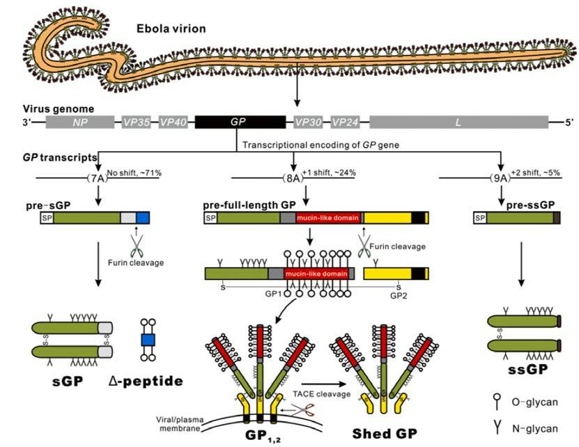
Ebolaviruses, a subclass of filoviruses belong to the genus Ebolavirus, causes Ebola virus disease characterized by a severe and often fatal hemorrhagic fever in humans and other mammals. As one of the structural proteins encoded by ebolaviruses, GP1, 2 glycoprotein is the only surface protein with highly glycosylated. GP1, 2 glycoproteins, also known as filoviral envelope glycoproteins, are transmembrane proteins that mediate cell association, a fusion of viral and cellular membranes, and then, mediate the entry of the viral core into the cytosol. Current vaccine candidate against ebolaviruses in various clinical and experimental trial stages are dominantly based on the GP1, 2 glycoproteins.
 Fig.1 Encoding strategy of ebolavirus GPs.1
Why N- and O-linked Glycoprofiling?
-
It is predicted that N-glycosylation sites and O-glycosylation sites of the GP1, 2 glycoproteins from ebolaviruses are respectively estimated to 17 and 30.
-
The number of glycosylation sites of the GP1, 2 glycoproteins is different from the other species.
-
Expression systems that used to produce GP1, 2 glycoproteins for research are different, which in turn impart different glycosylation patterns.
-
GP1, 2 N-glycans types alterations can affect its ability to bind to cell surface proteins as well as entry and affect the susceptibility to neutralizing antibodies.
Glycosylation and expression cells of GP1, 2 can affect protein immunogenicity, which consequently impacts the benefit of a given candidate vaccine. Furthermore, information on the glycosylation of the GP1, 2 glycoproteins is limited. Therefore, a direct comparison of glycan profiling of GP1, 2 glycoproteins is very necessary and much in demand.
Creative Biolabs has accumulated extensive experience in providing a full range of technical platforms to help our customers to promote their meaningful programs. For N- and O-linked glycoprofiling, we can offer a professional and perfect research strategy to you. Just feel free to contact us for more information and a detailed quote.
Reference
-
Ning, Yun-Jia, et al. "The roles of ebolavirus glycoproteins in viral pathogenesis." Virologica Sinica 32 (2017): 3-15. Distributed under Open Access license CC BY 4.0, without modification.
For Research Use Only.
Related Services:

 Fig.1 Encoding strategy of ebolavirus GPs.1


